. 2016 Feb 3;11(2):e0147254.
doi: 10.1371/journal.pone.0147254. eCollection 2016.

Identification of Human Islet Amyloid Polypeptide as a BACE2 Substrate

Affiliations

Identification of Human Islet Amyloid Polypeptide as a BACE2 Substrate

Ingrid C Rulifson et al. PLoS One. .

Abstract

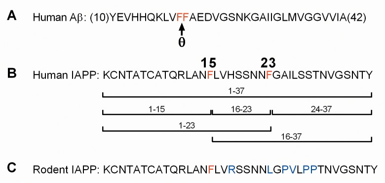
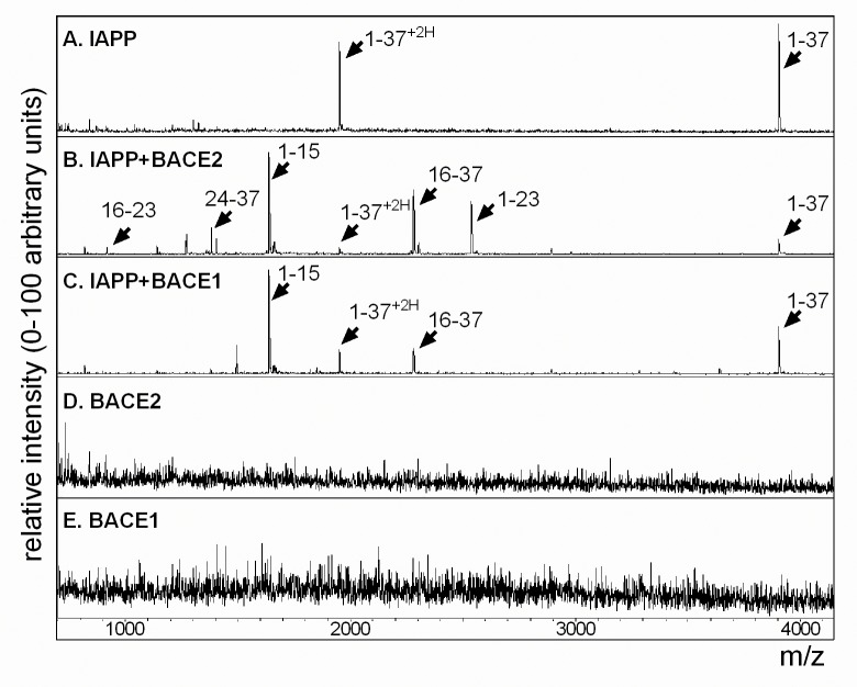
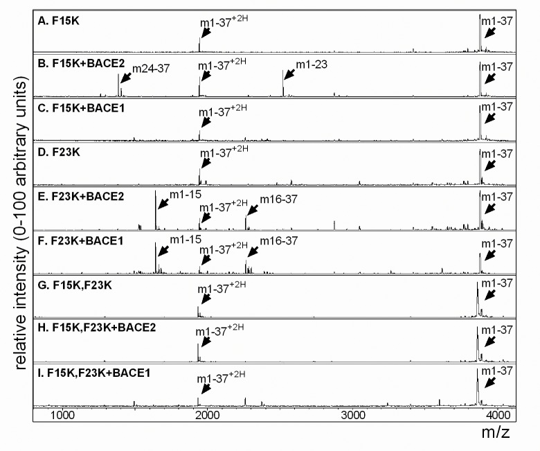
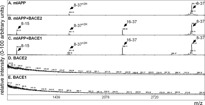
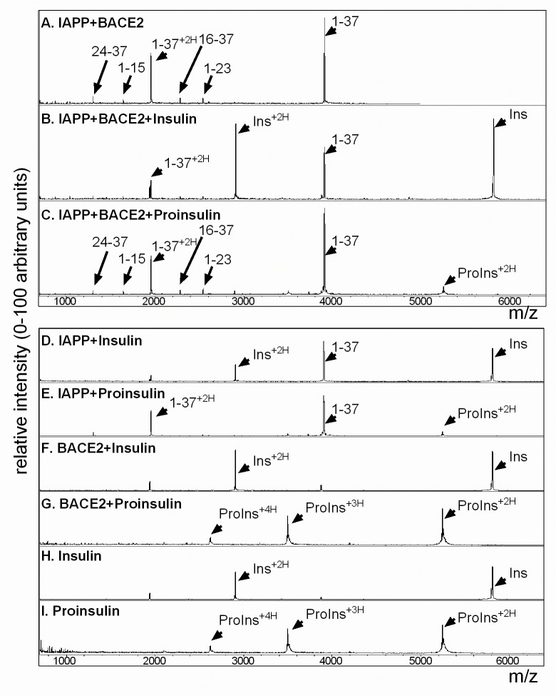
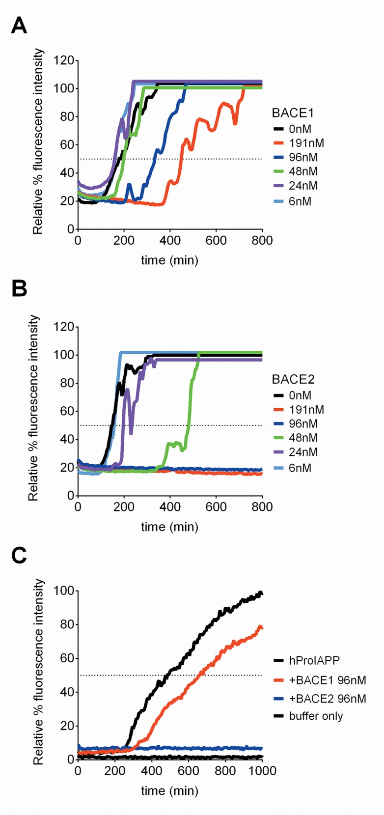
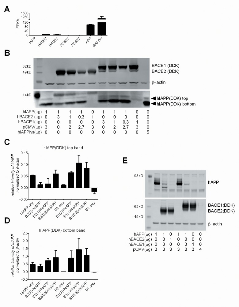
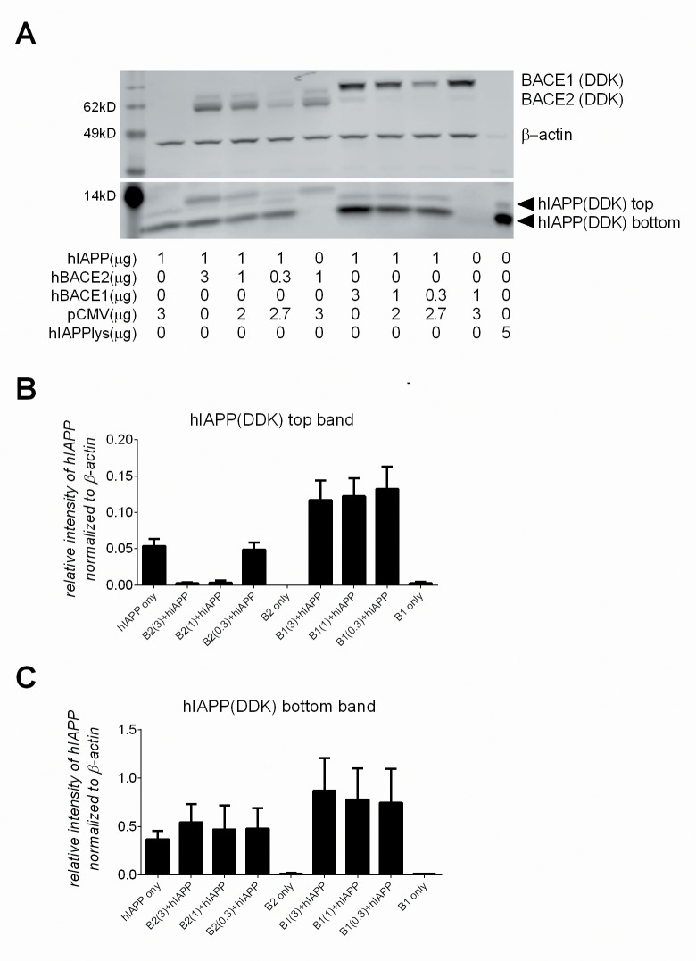
Pancreatic amyloid formation by islet amyloid polypeptide (IAPP) is a hallmark pathological feature of type 2 diabetes. IAPP is stored in the secretory granules of pancreatic beta-cells and co-secreted with insulin to maintain glucose homeostasis. IAPP is innocuous under homeostatic conditions but imbalances in production or processing of IAPP may result in homodimer formation leading to the rapid production of cytotoxic oligomers and amyloid fibrils. The consequence is beta-cell dysfunction and the accumulation of proteinaceous plaques in and around pancreatic islets. Beta-site APP-cleaving enzyme 2, BACE2, is an aspartyl protease commonly associated with BACE1, a related homolog responsible for amyloid processing in the brain and strongly implicated in Alzheimer's disease. Herein, we identify two distinct sites of the mature human IAPP sequence that are susceptible to BACE2-mediated proteolytic activity. The result of proteolysis is modulation of human IAPP fibrillation and human IAPP protein degradation. These results suggest a potential therapeutic role for BACE2 in type 2 diabetes-associated hyperamylinaemia.


Conflict of interest statement

Competing Interests: All authors are former or current Amgen employees. However this does not alter their adherence to PLOS ONE policies on sharing data and materials.

Figures

Fig 1
Fig 1. Comparison of human Aβ, human IAPP and rodent IAPP peptide sequences.
(A) The amino acid sequence of human Aβ (10–42); the described “theta” site (θ) is indicated by an arrow between phenylalanine residues 19 and 20 (in red). (B) The amino acid sequence of mature human IAPP; phenylalanine residues at positions 15 and 23 (in red). The potential peptide fragment species induced by BACE2-mediated cleavage are indicated: 1–37, 1–15, 16–23, 24–37, 1–23, and 16–37. (C) The amino acid sequence of mature rodent IAPP; the phenylalanine residue at position 15 (red) and residue differences between human and rodent sequences (blue) are highlighted.
Fig 2
Fig 2. Mass spectrometry of hIAPP confirms proteolytic cleavage by recombinant BACE2 at F15 and F23.
(A) MS analysis of hIAPP indicates a single protonated peak at m/z 3903 and a double protonated peak at m/z 1952. (B) Addition of recombinant BACE2 to hIAPP results in peaks at m/z 917; 16-LVHSSNNF-23, m/z 1382; 24-GAILSSTNVGSNTY-37, m/z 1639; 1-KCNTATCATQRLANF-15, m/z 2281; 16-LVHSSNNFGAILSSTNVGSNTY-37, and m/z 2537; 1-KCNTATCATQRLANFLVHSSNNF-23. (C) Addition of recombinant BACE1 indicates peaks only at m/z 1639 (1–15) and m/z 2281 (16–37), corresponding to cleavage only at F15. (D) Recombinant BACE2 alone and (E) recombinant BACE1 alone. All reactions were performed in 50mM NaOAc and 1M NaCl, pH 5 for 4 hours at 37°C. Peptide fragment sequences are detailed in Table 1.
Fig 3
Fig 3. hIAPP mutants block predicted digestion by BACE2 and BACE1.
(A) MS analysis of hIAPP F15K mutant peptide alone indicates a single protonated peak at m/z 3886 and a double protonated peak at m/z 1943. (B) Addition of recombinant BACE2 to mutant hIAPP F15K results in two new peaks at m/z 1382; 24-GAILSSTNVGSNTY-37, and m/z 2520; 1-KCNTATCATQRLANFLVHSSNNK-23. (C) Mutant hIAPP F15K is unchanged by the addition of recombinant BACE1. (D) Mutant hIAPP F23K. (E) Addition of recombinant BACE2 to mutant hIAPP F23K results in two new peaks at m/z 1639; 1-KCNTATCATQRLANK-15, and m/z 2263; 16-LVHSSNNFGAILSSTNVGSNTY-37. (F) Addition of recombinant BACE1 to mutant hIAPP F23K also results in two new peaks at m/z 1639; 1-KCNTATCATQRLANK-15, and m/z 2263; 16-LVHSSNNFGAILSSTNVGSNTY-37. (G) Double mutant, hIAPP F15K,F23K alone. Double mutant hIAPP F15K,F23K, is unchanged by the addition of recombinant BACE2 (H) or by the addition of recombinant BACE1 (I). All reactions were performed in 50mM NaOAc and 1M NaCl, pH 5 for 4 hours at 37°C. Peptide fragment sequences are detailed in Table 2.
Fig 4
Fig 4. BACE2 only clips mIAPP at phenylalanine 15.
(A) MS analysis of mIAPP peptide (8–37) indicates a single protonated peak at m/z 3200 and a double protonated peak at m/z 1602. (B) Addition of recombinant BACE2 to mIAPP peptide (8–37) results in two new peaks at m/z 920; 8-ATQRLANF-15, and m/z 2296; 16-LVRSSNNLGPVLPPTNVGSNTY-37. (C) Addition of recombinant BACE1 to mIAPP peptide (8–37) also results in two new peaks at m/z 920; 8-ATQRLANF-15, and m/z 2296; 16-LVRSSNNLGPVLPPTNVGSNTY-37. (D) Recombinant BACE2 alone and (E) recombinant BACE1 alone. All reactions were performed in 50mM NaOAc and 1M NaCl, pH 5 for 4 hours at 37°C. Peptide fragment sequences are detailed in Table 3.
Fig 5
Fig 5. BACE2-mediated proteolytic cleavage of hIAPP is blocked by insulin but not proinsulin.
(A) MS analysis of hIAPP incubated with recombinant BACE2; the predicted peaks are indicated: 1–37, 24–37, 1–15, 16–37 and 1–23. (B) Co-incubation with recombinant human insulin blocks BACE2-mediated digestion of hIAPP. (C) Co-incubation with recombinant human proinsulin does not prevent BACE2-mediated digestion of hIAPP. (D) hIAPP plus recombinant human insulin: Single protonated IAPP (m/z 3903) and double protonated IAPP (m/z 1952); single protonated insulin (m/z 5806) and double protonated insulin (m/z 2903). (E) hIAPP plus recombinant human proinsulin; single protonated IAPP (m/z 3903), double protonated IAPP (m/z 1952), double protonated proinsulin (m/z 5250), triple protonated proinsulin (m/z 3495), and quadruple protonated proinsulin (m/z 2625). (F) Recombinant insulin is unchanged by recombinant BACE2. (G) Recombinant proinsulin is unchanged by recombinant BACE2. (H) Recombinant insulin alone and (I) recombinant proinsulin alone. All reactions were performed in PBS, pH 7 for 4 hours at 37°C. Peptide fragment sequences detailed in Table 4.
Fig 6
Fig 6. BACE2 modulates hIAPP fibrillation.
The Thioflavin T graph demonstrates the time needed (x-axis) to reach 50% fibrillation (dotted black line) of human IAPP (solid black line) as depicted by relative % fluorescence intensity (y-axis). (A) Addition of recombinant BACE1 (6nM-191nM) slows the kinetics of hIAPP fibrillation in a dose-dependent manner. (B) Addition of recombinant BACE2 (6nM-191nM) blocks the kinetics of hIAPP fibrillation in a dose-dependent manner. (C) Addition of recombinant BACE1 (96nM, red line) slows the kinetics of hProIAPP fibrillation, whereas recombinant BACE2 (96nM, blue line) inhibits hProIAPP fibrillation. Lines indicate the average of the triplicate values for each condition.
Fig 7
Fig 7. BACE2 modulates human IAPP protein levels HEK293 cells.
(A) RNAseq FPKM values for basal expression of human IAPP, BACE2, BACE1, PCSK1, PCSK2 and APP in HEK293 cells. (B) HEK293 cells co-transfected with, or without, hIAPP plasmid DNA (1μg) and varying concentrations of hBACE2 plasmid DNA, hBACE1 plasmid DNA, or empty pCMV DNA, the latter to normalize DNA concentrations across conditions. A commercial source of Myc-DDK tagged hIAPP lysate (5 μg, last lane) was used to confirm identification of the hIAPP bands. The relative intensity of the top hIAPP band (C) and bottom hIAPP band (D) for each condition, as detected by anti-DDK and normalized to β-actin. Results are averaged from two separate experiments and the standard deviation is shown. (E) HEK293 cells co-transfected with hAPP, hBACE2, hBACE1 or empty pCMV DNA.
Fig 8
Fig 8. BACE2 modulates human IAPP protein levels in pancreatic beta-cells.
(A) βTC3 cells co-transfected with, or without, hIAPP plasmid DNA (1μg) and varying concentrations of hBACE2 plasmid DNA, hBACE1 plasmid DNA, or empty pCMV DNA, the latter to normalize DNA concentrations across conditions. A commercial source of Myc-DDK tagged hIAPP lysate (5 μg, last lane) was used to confirm identification of the hIAPP bands. The relative intensity of the top hIAPP band (B) and bottom hIAPP band (C) for each condition, as detected by anti-DDK and normalized to β-actin. Results are averaged from five separate experiments and the standard error is shown.
Fig 9
Fig 9. Evaluation of BACE inhibitors, Compound J and Compound 15, in B6.Cg-Lepob/J mice.
(A) The IC50s (μM) for Compound J and Compound 15. (B) Plasma clearance of Compound J and Compound 15 after a single oral gavage administration at 30mg/kg in 8–10 week old male B6.Cg-Lepob/J mice, n = 2 per time point. (C) Blood glucose levels before (0 minutes) and after an intraperitoneal glucose injection (10%/kg body weight) in B6.Cg-Lepob/J mice treated daily for 14 days with vehicle (black diamond, n = 14), Compound 15 (red circle, n = 13), Compound J (blue square, n = 13), or Exendin 4 (green triangle, n = 14). Asterisks indicate statistical significance based on 2-way ANOVA analysis, ** = 0.007, ***≤ 0.0007, **** < 0.0001. Target coverage analysis for BACE2 (D) and Tmem27 (E) on islets isolated from B6.Cg-Lepob/J mice treated with a single dose (30mg/kg, by oral gavage) of Compound J, Compound 15, or vehicle; n = 2 per group and islets were pooled for protein analysis. (F) One day before harvest, animals were injected with BrdU and pulsed for 24 hours prior to termination. Pancreas tissue was collected, stained for insulin and BrdU expression, and evaluated for several parameters to quantitate beta-cell proliferation. The image on the left shows a representative islet stained for insulin (Vulcan Red) and BrdU (DAB); the arrows point to Insulin+BrdU+ cells. The corresponding image demonstrates the morphometric analysis applied to quantitate proliferating beta-cells: individual insulin+ cells (green), insulin+BrdU+ cells (blue, indicated by arrows).

Similar articles

Cited by

  • Inside the Insulin Secretory Granule.
    Germanos M, Gao A, Taper M, Yau B, Kebede MA. Germanos M, et al. Metabolites. 2021 Aug 5;11(8):515. doi: 10.3390/metabo11080515. Metabolites. 2021. PMID: 34436456 Free PMC article. Review.
  • Physiological Functions of the β-Site Amyloid Precursor Protein Cleaving Enzyme 1 and 2.
    Yan R. Yan R. Front Mol Neurosci. 2017 Apr 19;10:97. doi: 10.3389/fnmol.2017.00097. eCollection 2017. Front Mol Neurosci. 2017. PMID: 28469554 Free PMC article. Review.
  • The β-Secretase BACE1 in Alzheimer's Disease.
    Hampel H, Vassar R, De Strooper B, Hardy J, Willem M, Singh N, Zhou J, Yan R, Vanmechelen E, De Vos A, Nisticò R, Corbo M, Imbimbo BP, Streffer J, Voytyuk I, Timmers M, Tahami Monfared AA, Irizarry M, Albala B, Koyama A, Watanabe N, Kimura T, Yarenis L, Lista S, Kramer L, Vergallo A. Hampel H, et al. Biol Psychiatry. 2021 Apr 15;89(8):745-756. doi: 10.1016/j.biopsych.2020.02.001. Epub 2020 Feb 13. Biol Psychiatry. 2021. PMID: 32223911 Free PMC article. Review.
  • Biological Mechanism-based Neurology and Psychiatry: A BACE1/2 and Downstream Pathway Model.
    Hampel H, Caruso G, Nisticò R, Piccioni G, Mercuri NB, Giorgi FS, Ferrarelli F, Lemercier P, Caraci F, Lista S, Vergallo A, Neurodegeneration Precision Medicine Initiative Npmi. Hampel H, et al. Curr Neuropharmacol. 2023;21(1):31-53. doi: 10.2174/1570159X19666211201095701. Curr Neuropharmacol. 2023. PMID: 34852743 Free PMC article.
  • The emerging role of β-secretases in cancer.
    Farris F, Matafora V, Bachi A. Farris F, et al. J Exp Clin Cancer Res. 2021 Apr 29;40(1):147. doi: 10.1186/s13046-021-01953-3. J Exp Clin Cancer Res. 2021. PMID: 33926496 Free PMC article. Review.

References

    1. Opie EL. Pathological Changes Affecting the Islands of Langerhans of the Pancreas. Journal Boston Society of Medical Sciences. 1900;4(10):251–60. - PMC - PubMed
    1. Wright AW. Hyaline Degeneration of the Islands of Langerhans in Non-Diabetics. The American journal of pathology. 1927;3(5):461–82 5. - PMC - PubMed
    1. Ehrlich JC, Ratner IM. Amyloidosis of the islets of Langerhans. A restudy of islet hyalin in diabetic and non-diabetic individuals. The American journal of pathology. 1961;38:49–59. - PMC - PubMed
    1. Pillay K, Govender P. Amylin uncovered: a review on the polypeptide responsible for type II diabetes. BioMed research international. 2013;2013:826706 10.1155/2013/826706 - DOI - PMC - PubMed
    1. Westermark P, Andersson A, Westermark GT. Islet amyloid polypeptide, islet amyloid, and diabetes mellitus. Physiological reviews. 2011;91(3):795–826. 10.1152/physrev.00042.2009 . - DOI - PubMed

MeSH terms

LinkOut - more resources