. 2016 Feb 3;12(2):e1005416.
doi: 10.1371/journal.ppat.1005416. eCollection 2016 Feb.

The Distribution of Prion Protein Allotypes Differs Between Sporadic and Iatrogenic Creutzfeldt-Jakob Disease Patients

Affiliations

The Distribution of Prion Protein Allotypes Differs Between Sporadic and Iatrogenic Creutzfeldt-Jakob Disease Patients

Roger A Moore et al. PLoS Pathog. .

Erratum in

Abstract

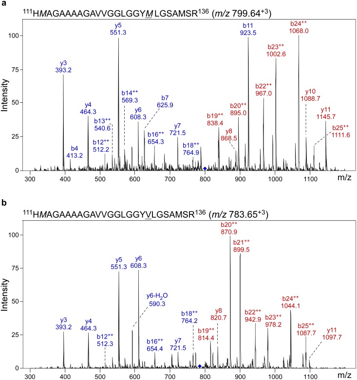
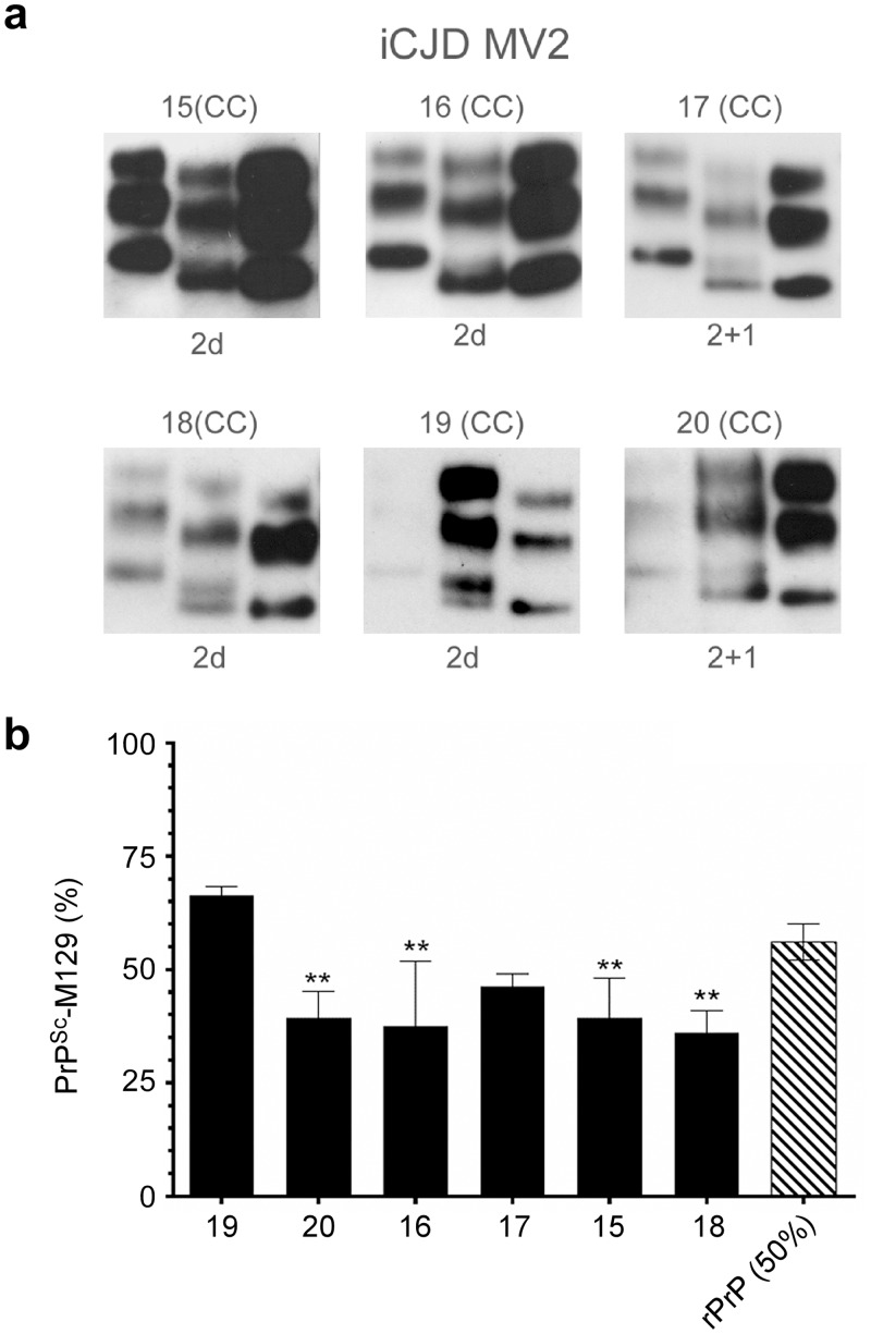
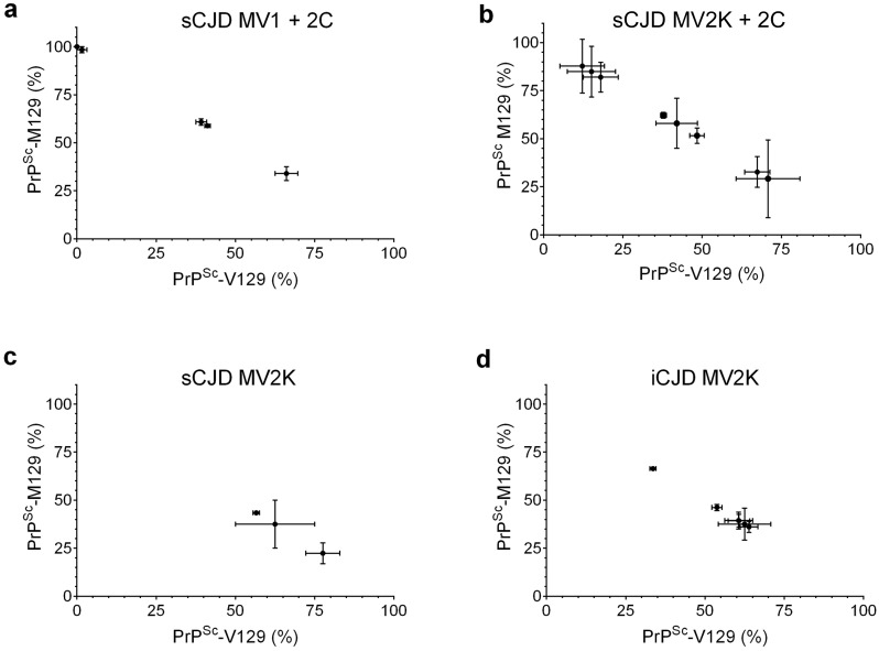
Sporadic Creutzfeldt-Jakob disease (sCJD) is the most prevalent of the human prion diseases, which are fatal and transmissible neurodegenerative diseases caused by the infectious prion protein (PrP(Sc)). The origin of sCJD is unknown, although the initiating event is thought to be the stochastic misfolding of endogenous prion protein (PrP(C)) into infectious PrP(Sc). By contrast, human growth hormone-associated cases of iatrogenic CJD (iCJD) in the United Kingdom (UK) are associated with exposure to an exogenous source of PrP(Sc). In both forms of CJD, heterozygosity at residue 129 for methionine (M) or valine (V) in the prion protein gene may affect disease phenotype, onset and progression. However, the relative contribution of each PrP(C) allotype to PrP(Sc) in heterozygous cases of CJD is unknown. Using mass spectrometry, we determined that the relative abundance of PrP(Sc) with M or V at residue 129 in brain specimens from MV cases of sCJD was highly variable. This result is consistent with PrP(C) containing an M or V at residue 129 having a similar propensity to misfold into PrP(Sc) thus causing sCJD. By contrast, PrP(Sc) with V at residue 129 predominated in the majority of the UK human growth hormone associated iCJD cases, consistent with exposure to infectious PrP(Sc) containing V at residue 129. In both types of CJD, the PrP(Sc) allotype ratio had no correlation with CJD type, age at clinical onset, or disease duration. Therefore, factors other than PrP(Sc) allotype abundance must influence the clinical progression and phenotype of heterozygous cases of CJD.


Conflict of interest statement

The authors have declared that no competing interests exist.

Figures

Fig 1
Fig 1. Methionine and valine polymorphism at residue 129 in human PrP can be distinguished by mass spectrometry.
Representative MS/MS spectra of human PrP peptide 111–136 with methionine (a) or valine (b) at residue 129 (underlined). The spectra are derived from a PK-treated, PTA-precipitated brain homogenate from sCJD case 10. The PrP111-136 MS/MS spectra were extracted from the raw data using the Data Analysis program v4.0 provided by Agilent. Ion scores of 130 and 127 were assigned by MASCOT for the M129 and V129 PrP111-136 peptides, respectively. Peaks were visualized using Scaffold v4.3 (Proteome Software, Portland, OR). The peptide sequence, charge state and observed m/z value for the MS1 ion are shown above each mass spectrum. Italicized methionine residues 112 and 129 in panel A and methionine 112 in panel B are oxidized in this representative example. Those fragment ions for which the m/z value changed depending upon the polymorphism at residue 129 are shown in red and those ions with m/z values unaffected by the changes at residue 129 are shown in blue. The m/z values shown are the experimentally observed values and deviate slightly from the values calculated in silico according to the mass accuracy of the instrument used in this study.
Fig 2
Fig 2. Representative neuropathology of heterozygous (MV) sCJD and iCJD patients analyzed.
(a) PrP immunohistochemistry performed on sections of cerebral cortex (CC) or cerebellar cortex (CbC) from sporadic CJD (sCJD) cases 1–5 of the MV1 molecular subtype and cases 6–14 of the MV2 molecular subtype. (b) PrP immunohistochemistry performed on sections of cerebral cortex (CC) from iatrogenic CJD (iCJD) cases 16–20 of the MV2 molecular subtype. Original magnifications were X400. The PrP antibody used was the mouse monoclonal antibody KG9 [34].
Fig 3
Fig 3. The relative abundance of PrPSc containing M at residue 129 is variable and independent of sCJD type in heterozygous (MV) sCJD patients.
(a) Molecular typing of PrPSc from the brain homogenate of heterozygous sCJD cases 1–14 by the method of Parchi et al. [33]. Each sample (middle lane) is shown flanked by Type 1 (left lane) and Type 2 (right lane) reference standards from sCJD MM1 and VV2 subtype cases, respectively. Protease-resistant PrP from all samples was of the A glycotype in which diglycosylated PrP ≤ monoglycosylated PrP [33]. Case number and brain region are indicated above each blot while PrPSc type is indicated below. The most informative exposure for the test and reference standard samples from a series of timed exposures is shown. CC = cerebral cortex; CbC = cerebellar cortex. (b) Relative abundance of PrPSc-M129 in MV1 and MV2 sCJD. Data are the percent mean ± SD of PrPSc-M129 (black bars) detected in the CC or CbC (samples 3, 7, 9, and 14 only) of sCJD patients. Each bar represents n = 3–4 technical replicates except for case 4 where n = 2. A stoichiometrically adjusted solution of 50% rHuPrP-M129 and 50% rHuPrP-V129 (rPrP (50%), hatched bar) was used as a control (n = 8). Statistical analysis was done using a 1-way ANOVA with Dunnett’s post-test using rPrP (50%) as the control sample. * p = 0.01 to 0.05, **p = 0.001 to 0.01, ***p = 0.0001 to 0.01, ****p <0.000.
Fig 4
Fig 4. The relative abundance of PrPSc containing M at residue 129 can vary between brain regions in heterozygous (MV) sCJD patients.
Data are the percent mean ± SD of PrPSc-M129 detected in the CbC (black bars) or CC (white bars). The case number is indicated under each pair of bars. Each bar represents n = 3–4 technical replicates except for the CC from cases 7 and 9 where n = 2. Statistical significance was determined using the unpaired Student’s t-test. *p = 0.01, **p = 0.002.
Fig 5
Fig 5. PrPSc containing M at residue 129 is underrepresented in most heterozygous (MV) iCJD patients.
(a) Molecular typing of PrPSc from the brain homogenate of heterozygous iCJD cases 15–20 by the method of Parchi et al. [33]. Each sample (middle lane) is shown flanked by Type 1 (left lane) and Type 2 (right lane) reference standards from sCJD MM1 and VV2 subtype cases, respectively. Case number and brain region are indicated above each blot while PrPSc type is indicated below. Protease-resistant PrP from all samples was of the A glycotype where diglycosylated PrP ≤ monoglycosylated PrP [33]. The most informative exposure for the test and reference standard samples from a series of timed exposures is shown. (b) Relative abundance of PrPSc-M129 in MV2 iCJD. Data are the percent mean ± SD of PrPSc-M129 (black bars) detected in the CC of iCJD patients. A stoichiometrically adjusted solution of 50% rHuPrP-M129 and 50% rHuPrP-V129 (rPrP (50%), hatched bar) was used as a control (n = 8). Each bar represents n = 3–4 technical replicates. Statistical analysis was done using a 1-way ANOVA with Dunnett’s post-test using rPrP (50%) as the control sample. **p = 0.001 to 0.01.
Fig 6
Fig 6. Variable PrPSc allotype ratios in sCJD neuropathological subtypes.
The percentage of PrPSc-M129 versus PrPSc-V129 is shown for neuropathological subtypes for which at least 2 cases were available for comparison. Each point represents the results from a single brain region from a single patient. For some patients, both CC and CbC were available for analysis. (a) sCJD MV1 + 2C, n = 4 cases, 5 samples total. (b) sCJD MV2K + 2C, n = 6 cases, 8 samples total. (c) sCJD MV2K, n = 2 cases, 3 samples total, (d) iCJD MV2K n = 6 cases. Using the unpaired Student’s t-test, the iCJD MV2K PrPSc allotype ratio differs significantly from both sCJD MV1 + 2C (p = 0.002) or sCJD MV2K + 2C (p = 0.02) but not from sCJD MV2K (p = 0.07).

Similar articles

Cited by

References

    1. Head MW, Ironside JW. Review: Creutzfeldt-Jakob disease: prion protein type, disease phenotype and agent strain. Neuropathol Appl Neurobiol. 2012;38: 296–310. 10.1111/j.1365-2990.2012.01265.x - DOI - PubMed
    1. Ghetti B, Piccardo P, Spillantini MG, Ichimiya Y, Porro M, Perini F, et al. Vascular variant of prion protein cerebral amyloidosis with tau-positive neurofibrillary tangles: the phenotype of the stop codon 145 mutation in PRNP. Proc Natl Acad Sci USA. 1996;93: 744–748. - PMC - PubMed
    1. Gambetti P, Puoti G, Zou WQ. Variably protease-sensitive prionopathy: a novel disease of the prion protein. J Mol Neurosci. 2011;45: 422–424. 10.1007/s12031-011-9543-1 - DOI - PubMed
    1. Puoti G, Bizzi A, Forloni G, Safar JG, Tagliavini F, Gambetti P. Sporadic human prion diseases: molecular insights and diagnosis. Lancet Neurol. 2012;11: 618–628. 10.1016/S1474-4422(12)70063-7 - DOI - PubMed
    1. Parchi P, Saverioni D. Molecular pathology, classification, and diagnosis of sporadic human prion disease variants. Folia Neuropathol. 2012;50: 20–45. - PubMed

Publication types

MeSH terms

Supplementary concepts