. 2016 May;148(1):92-101.
doi: 10.1111/imm.12590. Epub 2016 Mar 17.

Disorganization of the splenic microanatomy in ageing mice

Affiliations

Disorganization of the splenic microanatomy in ageing mice

Danielle Aw et al. Immunology. 2016 May.

Abstract

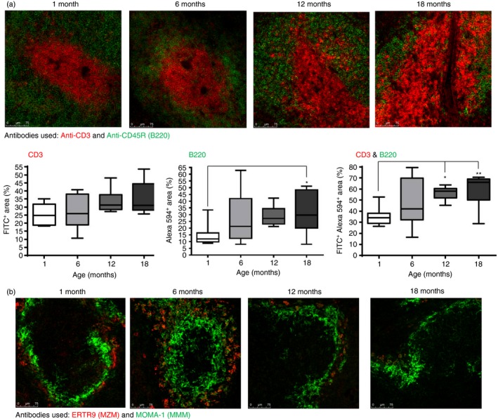
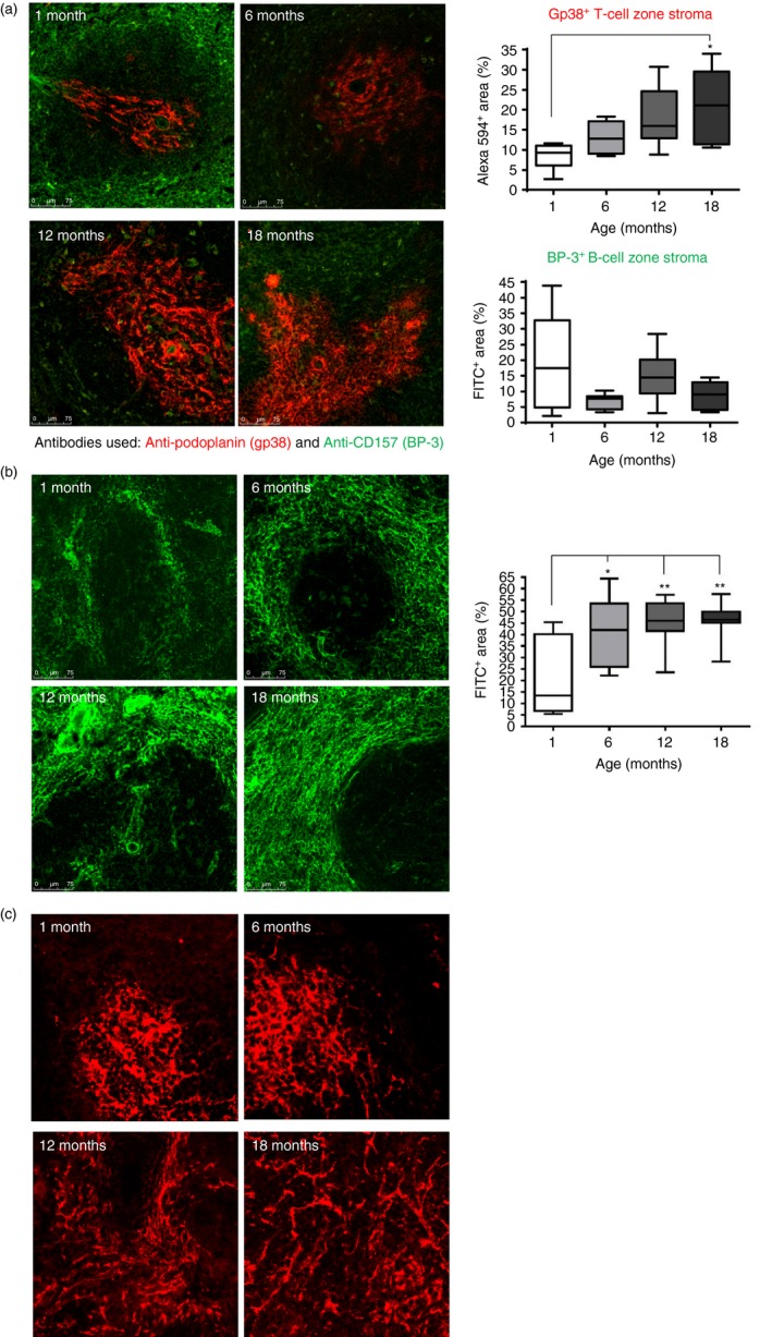
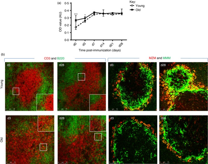
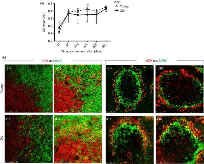
The precise mechanisms responsible for immunosenescence still remain to be determined, however, considering the evidence that disruption of the organization of primary and secondary lymphoid organs results in immunodeficiency, we propose that this could be involved in the decline of immune responses with age. Therefore, we investigated the integrity of the splenic microarchitecture in mice of increasing age and its reorganization following immune challenge in young and old mice. Several differences in the anatomy of the spleen with age in both the immune and stromal cells were observed. There is an age-related increase in the overall size of the white pulp, which occurs primarily within the T-cell zone and is mirrored by the enlargement of the T-cell stromal area, concurrent to the distinct boundary between T cells and B cells becoming less defined in older mice. In conjunction, there appears to be a loss of marginal zone macrophages, which is accompanied by an accumulation of fibroblasts in the spleens from older animals. Furthermore, whereas the reorganization of the white pulp is resolved after several days following antigenic challenge in young animals, it remains perturbed in older subjects. All these age-related changes within the spleen could potentially contribute to the age-dependent deficiencies in functional immunity.

Keywords: rodent; spleen and lymph nodes; stromal cells.


Figures

Figure 1
Figure 1
Changes of the splenic microenvironment with increasing age. (a) Splenic sections were stained with antibodies (anti‐CD3, anti‐B220) that recognized T cells (shown in green) and B cells (shown in red), respectively and the level of staining was assessed using imagej and portrayed as a percentage of the total area measured (FITC + Area refers to CD3, Alexa 594+ Area refers to B220 and FITC + Alexa 594+ Area refers to CD3 and B220). The data, displayed as graphs, are shown below the sections. (b) Splenic sections were stained with antibodies ERTR9 and anti‐MOMA‐1, which recognized marginal zone macrophages (MZM; in red) and marginal metallophilic macrophages (MMM; in green), respectively. The distinct demarcation of T and B cells within the splenic white pulp is lost with increasing age, which coincides with alterations in the marginal zone. Magnification ×100. Data representative of four experiments. *P < 0·05; **P < 0·01.
Figure 2
Figure 2
Stromal changes within spleens of young and old mice. (a) Sections were stained with the anti‐gp38 (podoplanin; in red), which detects the T‐cell stroma, and anti‐CD157 (BP‐3), which detects the B‐cell stroma, (shown in green). The level of staining was assessed by imageJ analysis (graphs on the right hand side) and revealed an increase in the T‐cell stromal area. Magnification ×100. (b) Staining for the fibroblastic reticular cell (FRC) with ERTR7 antibody revealed an increased deposition of FDC around the marginal zone in the splenic sections from older mice. Magnification ×100. (c) Staining for follicular dendritic cells (FDC) using anti‐FDC‐M2 revealed that the network of FDC appears more dispersed in the splenic sections from older mice Magnification ×200. Isotype controls revealed no staining (data not shown). Data representative of four experiments. *P < 0·05; **P < 0·01.
Figure 3
Figure 3
Architectural changes in the splenic microenvironment following immunization with a T‐independent antigen. (a) Blood was taken from young (1 month) and old (18 months) mice at days 0, 3, 7, 14, 21 and 28 after immunization with trintriphenyl (TNP) –Ficoll and the specific anti‐IgM TNP–Ficoll response was determined by ELISA. Both young and old mice show a similar increase in the IgM response after immunization, although the level of the anti‐IgM TNP–Ficoll antibody is initially higher at days 0 and 3 in older animals. Six mice were used for each time‐point. *P < 0·05; ***P < 0·001. Data representative of three experiments. (b) Splenic sections of young and old mice were stained to detect T (red) and B (green) cells and marginal zone macrophages (MZM; red) and marginal metallophilic macrophages (MMM; green). Following immunization, the distinct demarcation of the T‐ and B‐cell zone of both age groups is disrupted (day 3), which is resolved by day 28 in young mice, but is still disorganized in the spleen of old mice. Magnification ×100. Insert of an area highlighted by a white box is shown representing a higher magnification (×200). In the spleen of young mice MMM and MZM encircle the marginal zone, which is incomplete, despite the appearance of MZM, in old animals. Magnification ×100. Isotype controls revealed no staining (data not shown). Data representative of four experiments.
Figure 4
Figure 4
Alterations in the splenic microenvironment following immunization with a T‐cell‐dependent antigen. (a) Young (1 month) and old (18 months) mice were immunized with trinitrophenyl–keyhole limpet haemocyanin (TNPKLH) at day 0 and day 28, and blood was taken from young (1 month) and old (18 months) mice at days; 0, 7, 14, 21, 28 and 35 after immunization. Level of anti‐IgG TNPKLH antibody increases after immunization in both age groups; young mice show a higher level at day 21. Although older mice showed a higher level before immunization. Six mice were used for each time‐point. *P < 0·05; **P < 0·01. Data representative of three experiments. (b) Splenic sections of young and old mice were stained with anti‐CD3 (green) and anti‐B220 (red) and revealed disruption of the T‐cell and B‐cell zone in both age groups, although it was more apparent in the sections from older mice. Sections stained with marginal zone macrophages (MZM; red) and marginal metallophilic macrophages (MMM; green) reveals that in the spleens of young mice MMM and MZM encircle the marginal zone, which is incomplete, despite the appearance of MZM, in old animals. Magnification ×100. Isotype controls revealed no staining (data not shown). Data representative of four experiments.

References

    1. Aw D, Silva AB, Palmer DB. Immunosenescence: emerging challenges for an ageing population. Immunology 2007; 120:435–46. - PMC - PubMed
    1. Haynes L, Swain SL. Why aging T cells fail: implications for vaccination. Immunity 2006; 24:663–6. - PMC - PubMed
    1. Pawelec G, Akbar A, Caruso C, Solana R, Grubeck‐Loebenstein B, Wikby A. Human immunosenescence: is it infectious? Immunol Rev 2005; 205:257–68. - PubMed
    1. Chen WH, Kozlovsky BF, Effros RB, Grubeck‐Loebenstein B, Edelman R, Sztein MB. Vaccination in the elderly: an immunological perspective. Trends Immunol 2009; 30:351–9. - PMC - PubMed
    1. Gardner ID. The effect of aging on susceptibility to infection. Rev Infect Dis 1980; 2:801–10. - PubMed

Publication types

LinkOut - more resources