

An Optical Section-Assisted In Vivo Rabbit Model for Capsular Bend and Posterior Capsule Opacification Investigation
- PMID: 26840405
- PMCID: PMC4739694
- DOI: 10.1371/journal.pone.0148553
An Optical Section-Assisted In Vivo Rabbit Model for Capsular Bend and Posterior Capsule Opacification Investigation
Abstract
Purpose: To establish an optical section-assisted in vivo rabbit model for capsular bend and posterior capsule opacification (PCO) investigation.
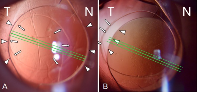
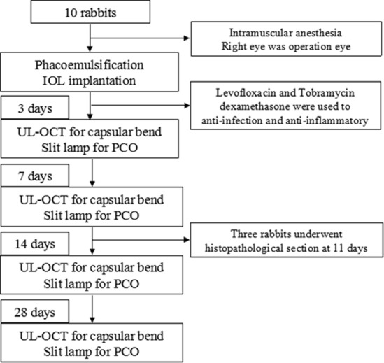
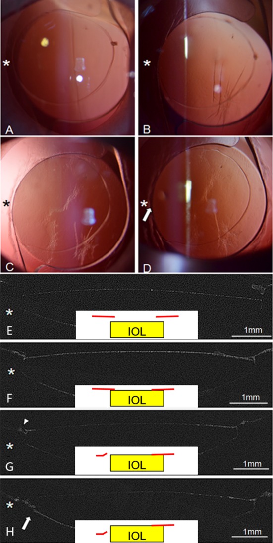
Methods: A total of 10 rabbits underwent phacoemulsification surgery and intraocular lens (IOL) implantation. On the basis of the relationship between the anterior capsule and IOL, the rabbits were divided into complete overlap and incomplete overlap groups, in which six and four rabbits were included, respectively. The capsular bend optical sections were assessed using ultra-long scan depth optical coherence tomography (UL-OCT), and posterior capsule opacification was evaluated with slit lamp on postoperative day 3, 7, 14, and 28. In addition, histopathological section was used to verify the accuracy of capsular bend type captured by OCT in three rabbits.
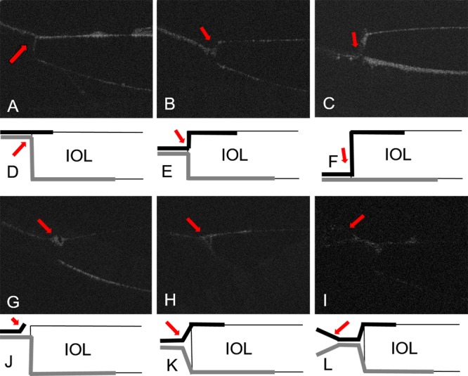
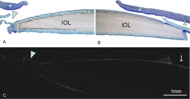
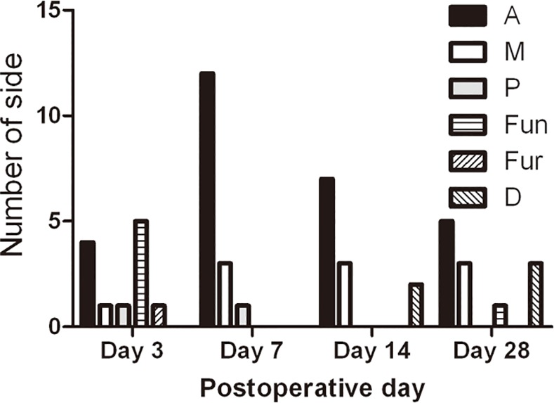
Results: Based on the special animal model, six capsular bend types were observed, namely, anterior (A), middle (M), posterior (P), detachment (D), funnel (Fun) and furcate adhesion (Fur). On day 3, capsular bend began to form. On 14 days, the capsular bends were comprised of A, M and D types, which were almost maintained until day 28. Histopathological section findings were consistent with optical sectioning results. In the incomplete and complete groups, the earliest PCO within the optical zone were on day 7 and 28, respectively. The incomplete group exhibited higher incidence and faster PCO on day 7 (p = 0.038) and 14 (p = 0.002).
Conclusions: This animal model not only mimics capsular bend evolution and PCO processes but also produces OCT optical section images equivalent to and more repeatable than histopathology, thereby providing a promising method for the further investigations of PCO.
Conflict of interest statement
Figures

Similar articles
-
Analysis of the capsular bend in posterior capsular opacification using anterior segment optical coherence tomography.Int Ophthalmol. 2023 Dec;43(12):4945-4958. doi: 10.1007/s10792-023-02897-7. Epub 2023 Oct 28. Int Ophthalmol. 2023. PMID: 37897540 Free PMC article.
-
Relationship of Posterior Capsular Opacification and Capsular Bend Type Investigation Based on Swept-source Optical Coherence Tomography.Curr Eye Res. 2020 Jan;45(1):17-23. doi: 10.1080/02713683.2019.1645183. Epub 2019 Jul 29. Curr Eye Res. 2020. PMID: 31348676
-
Capsular adhesion to intraocular lens in highly myopic eyes evaluated in vivo using ultralong-scan-depth optical coherence tomography.Am J Ophthalmol. 2013 Mar;155(3):484-491.e1. doi: 10.1016/j.ajo.2012.08.019. Epub 2012 Dec 4. Am J Ophthalmol. 2013. PMID: 23218694
-
Posterior capsule opacification.Curr Opin Ophthalmol. 2006 Feb;17(1):45-53. doi: 10.1097/01.icu.0000193074.24746.e6. Curr Opin Ophthalmol. 2006. PMID: 16436924 Review.
-
Prevention of posterior capsular opacification.Exp Eye Res. 2015 Jul;136:100-15. doi: 10.1016/j.exer.2015.03.011. Epub 2015 Mar 14. Exp Eye Res. 2015. PMID: 25783492 Review.
Cited by
-
Application of Spectral Domain Optical Coherence Tomography to Objectively Evaluate Posterior Capsular Opacity In Vivo.J Ophthalmol. 2018 Dec 23;2018:5461784. doi: 10.1155/2018/5461784. eCollection 2018. J Ophthalmol. 2018. PMID: 30671258 Free PMC article.
-
Factors Affecting Posterior Capsule Opacification in the Development of Intraocular Lens Materials.Pharmaceutics. 2021 Jun 10;13(6):860. doi: 10.3390/pharmaceutics13060860. Pharmaceutics. 2021. PMID: 34200928 Free PMC article. Review.
-
Time Course of Lens Epithelial Cell Behavior in Rabbit Eyes following Lens Extraction and Implantation of Intraocular Lens.J Ophthalmol. 2021 Jan 16;2021:6659838. doi: 10.1155/2021/6659838. eCollection 2021. J Ophthalmol. 2021. PMID: 33510905 Free PMC article.
-
Analysis of the capsular bend in posterior capsular opacification using anterior segment optical coherence tomography.Int Ophthalmol. 2023 Dec;43(12):4945-4958. doi: 10.1007/s10792-023-02897-7. Epub 2023 Oct 28. Int Ophthalmol. 2023. PMID: 37897540 Free PMC article.
References
-
- Knight-Nanan D, O'Keefe M, Bowell R. Outcome and complications of intraocular lenses in children with cataract. Journal of cataract and refractive surgery. 1996;22(6):730–6. - PubMed
-
- Sacu S, Findl O, Menapace R, Buehl W. Influence of optic edge design, optic material, and haptic design on capsular bend configuration. Journal of cataract and refractive surgery. 2005;31(10):1888–94. - PubMed
-
- Nishi O, Yamamoto N, Nishi K, Nishi Y. Contact inhibition of migrating lens epithelial cells at the capsular bend created by a sharp-edged intraocular lens after cataract surgery. Journal of cataract and refractive surgery. 2007;33(6):1065–70. - PubMed
-
- Nishi O, Nishi K, Sakanishi K. Inhibition of migrating lens epithelial cells at the capsular bend created by the rectangular optic edge of a posterior chamber intraocular lens. Ophthalmic surgery and lasers. 1998;29(7):587–94. - PubMed
-
- Nishi O, Nishi K, Osakabe Y. Effect of intraocular lenses on preventing posterior capsule opacification: design versus material. Journal of cataract and refractive surgery. 2004;30(10):2170–6. - PubMed
Publication types
MeSH terms
LinkOut - more resources
Full Text Sources
Other Literature Sources
