. 2016 Feb 3;11(2):e0147907.
doi: 10.1371/journal.pone.0147907. eCollection 2016.

Neisseria meningitidis Translation Elongation Factor P and Its Active-Site Arginine Residue Are Essential for Cell Viability

Affiliations

Neisseria meningitidis Translation Elongation Factor P and Its Active-Site Arginine Residue Are Essential for Cell Viability

Tatsuo Yanagisawa et al. PLoS One. .

Abstract

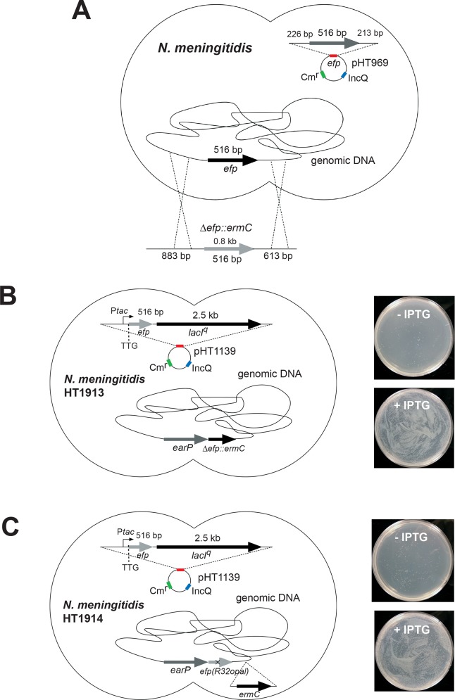
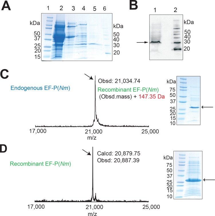
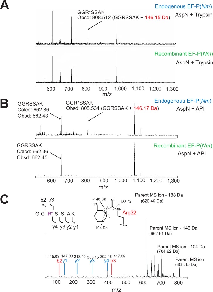
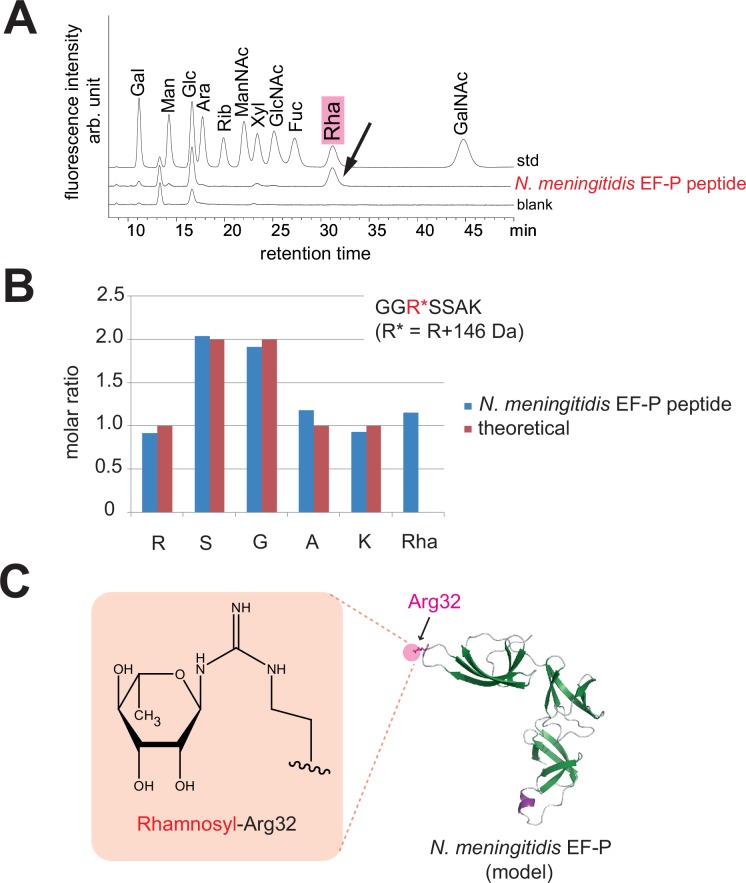
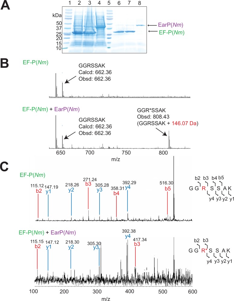
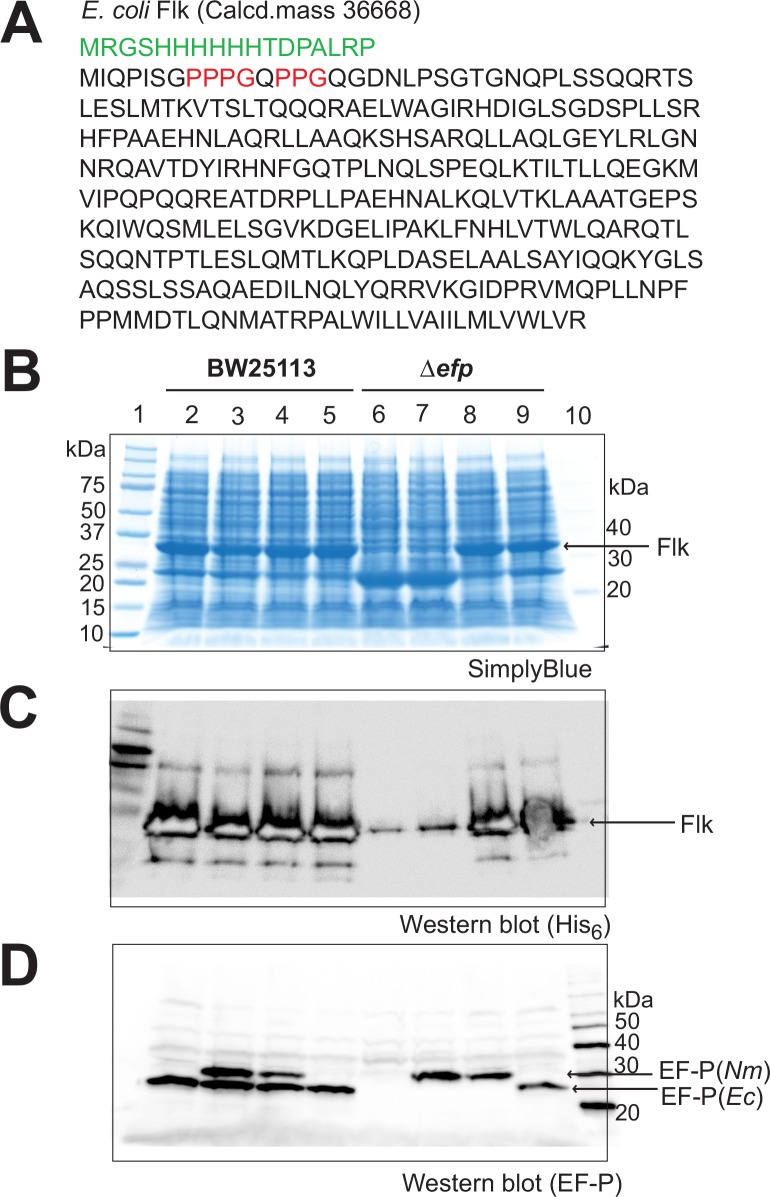
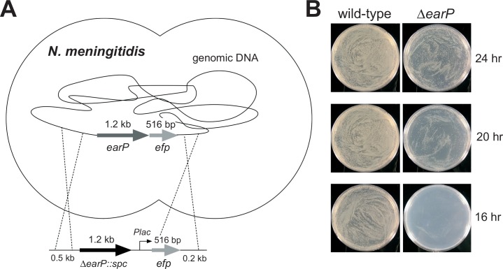
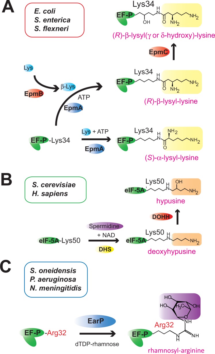
Translation elongation factor P (EF-P), a ubiquitous protein over the entire range of bacterial species, rescues ribosomal stalling at consecutive prolines in proteins. In Escherichia coli and Salmonella enterica, the post-translational β-lysyl modification of Lys34 of EF-P is important for the EF-P activity. The β-lysyl EF-P modification pathway is conserved among only 26-28% of bacteria. Recently, it was found that the Shewanella oneidensis and Pseudomonas aeruginosa EF-P proteins, containing an Arg residue at position 32, are modified with rhamnose, which is a novel post-translational modification. In these bacteria, EF-P and its Arg modification are both dispensable for cell viability, similar to the E. coli and S. enterica EF-P proteins and their Lys34 modification. However, in the present study, we found that EF-P and Arg32 are essential for the viability of the human pathogen, Neisseria meningitidis. We therefore analyzed the modification of Arg32 in the N. meningitidis EF-P protein, and identified the same rhamnosyl modification as in the S. oneidensis and P. aeruginosa EF-P proteins. N. meningitidis also has the orthologue of the rhamnosyl modification enzyme (EarP) from S. oneidensis and P. aeruginosa. Therefore, EarP should be a promising target for antibacterial drug development specifically against N. meningitidis. The pair of genes encoding N. meningitidis EF-P and EarP suppressed the slow-growth phenotype of the EF-P-deficient mutant of E. coli, indicating that the activity of N. meningitidis rhamnosyl-EF-P for rescuing the stalled ribosomes at proline stretches is similar to that of E. coli β-lysyl-EF-P. The possible reasons for the unique requirement of rhamnosyl-EF-P for N. meningitidis cells are that more proline stretch-containing proteins are essential and/or the basal ribosomal activity to synthesize proline stretch-containing proteins in the absence of EF-P is lower in this bacterium than in others.


Conflict of interest statement

Competing Interests: The authors have declared that no competing interests exist.

Figures

Fig 1
Fig 1. Strategies for efp deletion from the N. meningitidis genome.
(A) The efp allele in the N. meningitidis genome can be disrupted, but only in the presence of a plasmid containing the wild-type N. meningitidis efp gene. (B, C) The IncQ plasmid pHT1139, containing Ptac-TTG-efp-lacIq, was transformed into N. meningitidis H44/76 cells. Subsequently, the DNA fragment bearing the erythromycin resistance gene (ermC) or the earP-efp(R32opal) gene was introduced into the efp allele within the N. meningitidis H44/76/pHT1139 genome, to obtain the N. meningitidis strains HT1913/pHT1139 (B, left) and HT1914/pHT1139 (C, left), respectively. In these strains, the efp gene expression can be controlled by IPTG, and EF-P can be inducibly produced in the presence of IPTG. (B, C, right) Growth of the N. meningitidis HT1913/pHT1139 and HT1914/pHT1139 cells, with and without IPTG. Both of the N. meningitidis cells lack the efp gene in the genome, but contain an inducible copy of the efp gene in the IncQ plasmid.
Fig 2
Fig 2. Purification and MS analysis of N. meningitidis EF-P(Nm).
Proteins in each purification step were analyzed by 10–20% SDS-PAGE and stained with SimplyBlue SafeStain. (A) Lane 1, molecular mass standards; lane 2, N. meningitidis crude cell extract; lane 3, after DEAE-Sephacel column; lane 4, after HiTrap Q HP column; lane 5, endogenous EF-P(Nm) purified on a HiTrap Butyl HP column; lane 6, MagicMark molecular mass standards (Life Technologies). (B) The polyclonal antibody against EF-P(Ec) cross-reacts with EF-P(Nm). The EF-P proteins in the N. meningitidis cell extracts and the column chromatography fractions were monitored by western blotting with the antibody. Lane 1, N. meningitidis crude cell extract; lane 2, MagicMark molecular mass standards. The arrow designates EF-P(Nm). (C) MALDI-TOF MS and SDS-PAGE of the endogenous EF-P(Nm), shown in the left and right panels, respectively. (D) MALDI-TOF MS and SDS-PAGE of the recombinant EF-P(Nm), shown in the left and right panels, respectively.
Fig 3
Fig 3. PMF and MS/MS analyses of N. meningitidis endogenous EF-P.
(A, B) The endogenous and recombinant EF-P(Nm) proteins were digested with AspN and trypsin (A) or with AspN and API (B), and were subjected to the PMF analysis. The peptide “GGR*SSAK” (R* represents modified Arg32) of the endogenous EF-P(Nm) was larger by 146 Da (obsd: 808.512 [M+H]+ and 808.534 [M+H]+), than the calculated mass of the peptide “GGRSSAK” obtained from the recombinant EF-P(Nm) (calcd: 662.36 [M+H]+, obsd: 662.43 [M+H]+). (C) MS/MS spectrum of the AspN and trypsin-digested endogenous EF-P(Nm). The sequence can be read from the annotated b (blue) or y (red) ion series; the b2, b3, y1, y2, y3, and y4 ions were observed. The parent ion (obsd: 808.454 [M+H]+), and parent ion –104 Da (obsd: 704.624 [M+H]+), parent ion –146 Da (obsd: 662.61 [M+H]+), and parent ion –188 Da (obsd: 620 462 [M+H]+) generated by the degradation of the modified arginine residue were also observed. Possible degradation sites of the modified arginine are represented by dotted lines.
Fig 4
Fig 4. Analysis of the sugar and amino acid components in the modified peptide of N. meningitidis endogenous EF-P.
(A) Analysis of the sugar composition in the endogenous EF-P(Nm) peptide, GGR*SSAK. The peptide was hydrolyzed to free amino acids and other components. The samples were derivatized with ABEE, and were subjected to the UHPLC analysis, using 11 monosaccharides as standards (std). A blank sample including deionized water was also loaded (blank). Gal, galactose; Man, mannose; Glc, glucose; Ara, arabinose; Rib, ribose; ManNAc, N-acetylmannosamine; Xyl, xylose; GlcNAc, N-acetylglucosamine; Fuc, fucose; Rha, rhamnose; GalNAc, N-acetylgalactosamine. (B) Quantitative analysis of the amino acid and rhamnose components in the EF-P(Nm) peptide. (C) Rhamnosyl–Arg32 and a 3D structural model of N. meningitidis EF-P.
Fig 5
Fig 5. Rhamnosyl modification of the recombinant EF-P(Nm) by EarP(Nm).
(A) Coexpression of EF-P(Nm) with EarP(Nm) in E. coli cells. Lane 1, molecular mass standards; lane 2, crude extract of E. coli cells producing EF-P(Nm); lane 3, crude extract of E. coli cells producing EF-P(Nm) and EarP(Nm); lane 4, crude extract of E. coli cells producing EarP(Nm); lane 5, molecular mass standards; lane 6, the recombinant EF-P(Nm) purified from the cells producing EF-P(Nm); lane 7, the recombinant EF-P(Nm) purified from the cells producing EF-P(Nm) and EarP(Nm); lane 8, purified EarP(Nm). (B) PMF analysis of the modified and unmodified EF-P(Nm). The recombinant EF-P(Nm) proteins were purified from the cells producing EF-P(Nm), with or without EarP(Nm). After digestion with AspN and API, the PMF analysis of the peptides was performed. (C) MS/MS analyses of the recombinant EF-P(Nm) and the recombinant EF-P(Nm) modified with EarP(Nm). After digestion with AspN and API, the EF-P(Nm) peptides with masses of 662.35 Da (GGRSSAK) and 808.4 Da (GGR*SSAK, R* designates the modified Arg32) were subjected to the MS/MS analysis. The sequence can be read from the annotated b (blue) or y (red) ion series; the b2, b3, b4, b5, y1, y2, y3, and y4 ions from the peptide “GGRSSAK” and the b2, b3, y1, y2, y3, and y4 ions from the peptide “GGR*SSAK” were observed.
Fig 6
Fig 6. Restoration of production of the E. coli Flk protein, containing proline stretches, in efp-deficient cells.
(A) Amino acid sequence of the E. coli Flk protein. Proline stretches in Flk are colored red. (B) Plasmids pMW119, pMW-NmE (a pMW119-derived plasmid for expressing EF-P(Nm)), pMW-NmED (a pMW119-derived plasmid for expressing EF-P(Nm) and EarP(Nm)), and pMW-EcEGY (a pMW119-derived plasmid for expressing E. coli EF-P, EpmA, and EpmB) were cotransformed with the Flk plasmid into E. coli BW25113 and JW4107 (BW25113 Δefp::kan) cells. Whole-cell proteins were subjected to SDS-PAGE, and analyzed for the production of the full-length Flk protein. Lane 1: molecular mass standards; lane 2: BW25113/pMW119; lane 3: BW25113/pMW-NmE; lane 4: BW25113/pMW-NmED; lane 5: BW25113/pMW-EcEGY; lane 6: JW4107/pMW119; lane 7: JW4107/pMW-NmE; lane 8: JW4107/pMW-NmED; lane 9: JW4107/pMW-EcEGY; lane 10, MagicMark molecular mass standards. Expression of the chloramphenicol acetyltransferase gene in the Flk plasmid was also observed (23 kDa). (C) The Flk protein was recognized by an anti-His6-antibody. (D) Western blotting using a polyclonal antibody against E. coli EF-P. The expressed EF-P(Nm) cross-reacted with the anti-EF-P(Ec) antibody.
Fig 7
Fig 7. The earP gene is important, but not essential, for N. meningitidis viability.
(A) Strategy for the earP deletion in N. meningitidis. (B) Growth of N. meningitidis HT1125 wild-type (left) and the earP-deleted cells (right).
Fig 8
Fig 8. Post-translational modifications of EF-P and eIF5A.
(A) β-Lysylation and subsequent hydroxylation of EF-P(Lys34) in E. coli, S. enterica, and S. flexneri. EpmB catalyzes the conversion of (S)-lysine (L-lysine) to (R)-β-lysine, and EpmA mainly uses (R)-β-lysine as the substrate for the production of (R)-β-lysyl–EF-P. β-Lys represents (R)-β-lysine. (B) Deoxyhypusine modification and subsequent hydroxylation of eIF5A(Lys50) in H. sapiens and S. cerevisiae. (C) Rhamnosyl modification of EF-P(Arg32) in S. oneidensis, P. aeruginosa, and N. meningitidis. The DUF2331 superfamily proteins (EarP) that catalyze the rhamnosyl modification of EF-P(Arg32) are encoded in the genomes of N. meningitidis, N. gonorrhoeae (accession code: AAW89632), B. pertussis (accession code: CAE42291), B. cepacia (accession code: AIO49016), K. kingae (accession code: EIC14610), R. solanacearum (accession code: CBJ51687), P. aeruginosa (accession code: WP_034078567), P. fluorescens (accession code: AIG05734), and A. hydrophila (accession code: ABK36714), but are not encoded in those of B. burgdorferi, T. thermophilus, or D. radiodurans.

Similar articles

Cited by

References

    1. Pavlov MY, Watts RE, Tan Z, Cornish VW, Ehrenberg M, Forster AC. Slow peptide bond formation by proline and other N-alkylamino acids in translation. Proc Natl Acad Sci USA. 2009; 106: 50–54. 10.1073/pnas.0809211106 - DOI - PMC - PubMed
    1. Tanner DR, Cariello DA, Woolstenhulme CJ, Broadbent MA, Buskirk AR. Genetic identification of nascent peptides that induce ribosome stalling. J Biol Chem. 2009; 284: 34809–34818. 10.1074/jbc.M109.039040 - DOI - PMC - PubMed
    1. Doerfel LK, Wohlgemuth I, Kothe C, Peske F, Urlaub H, Rodnina MV. EF-P is essential for rapid synthesis of proteins containing consecutive proline residues. Science 2013; 339: 85–88. 10.1126/science.1229017 - DOI - PubMed
    1. Ude S, Lassak J, Starosta AL, Kraxenberger T, Wilson DN, Jung K. Translation elongation factor EF-P alleviates ribosome stalling at polyproline stretches. Science 2013; 339: 82–85. 10.1126/science.1228985 - DOI - PubMed
    1. Woolstenhulme CJ, Parajuli S, Healey DW, Valverde DP, Petersen EN, Starosta AL, et al. Nascent peptides that block protein synthesis in bacteria. Proc Natl Acad Sci USA. 2013; 110: E878–887. 10.1073/pnas.1219536110 - DOI - PMC - PubMed

Publication types

MeSH terms

LinkOut - more resources