. 2016 Jul 15;194(2):156-68.
doi: 10.1164/rccm.201507-1414OC.

Rapid Expansion of Human Epithelial Stem Cells Suitable for Airway Tissue Engineering

Affiliations

Rapid Expansion of Human Epithelial Stem Cells Suitable for Airway Tissue Engineering

Colin R Butler et al. Am J Respir Crit Care Med. .

Abstract

Rationale: Stem cell-based tracheal replacement represents an emerging therapeutic option for patients with otherwise untreatable airway diseases including long-segment congenital tracheal stenosis and upper airway tumors. Clinical experience demonstrates that restoration of mucociliary clearance in the lungs after transplantation of tissue-engineered grafts is critical, with preclinical studies showing that seeding scaffolds with autologous mucosa improves regeneration. High epithelial cell-seeding densities are required in regenerative medicine, and existing techniques are inadequate to achieve coverage of clinically suitable grafts.

Objectives: To define a scalable cell culture system to deliver airway epithelium to clinical grafts.

Methods: Human respiratory epithelial cells derived from endobronchial biopsies were cultured using a combination of mitotically inactivated fibroblasts and Rho-associated protein kinase (ROCK) inhibition using Y-27632 (3T3+Y). Cells were analyzed by immunofluorescence, quantitative polymerase chain reaction, and flow cytometry to assess airway stem cell marker expression. Karyotyping and multiplex ligation-dependent probe amplification were performed to assess cell safety. Differentiation capacity was tested in three-dimensional tracheospheres, organotypic cultures, air-liquid interface cultures, and an in vivo tracheal xenograft model. Ciliary function was assessed in air-liquid interface cultures.

Measurements and main results: 3T3-J2 feeder cells and ROCK inhibition allowed rapid expansion of airway basal cells. These cells were capable of multipotent differentiation in vitro, generating both ciliated and goblet cell lineages. Cilia were functional with normal beat frequency and pattern. Cultured cells repopulated tracheal scaffolds in a heterotopic transplantation xenograft model.

Conclusions: Our method generates large numbers of functional airway basal epithelial cells with the efficiency demanded by clinical transplantation, suggesting its suitability for use in tracheal reconstruction.

Keywords: adult stem cells; epithelium; respiratory mucosa; tissue engineering; trachea.


Figures

Figure 1.
Figure 1.
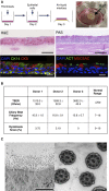
Isolation of autologous human basal epithelial cells from tracheobronchial biopsies. (A) Schematic representation of epithelial cell culture pipeline from bronchoscopic biopsy explants. (B) Bright-field image of biopsy outgrowth (scale bar, 20 μm). (C) Table summarizing flow cytometric analysis of the proportion of basal cell marker–expressing cells grown from biopsies in bronchial epithelial growth medium (BEGM) (n = 5 biopsies). (D) Decellularized human tracheal scaffolds seeded with expanded CK5+ airway epithelial cells (green) show poor coverage at seeding densities of less than 1 × 106 cells/cm2 (counterstained with 4′,6-diamidino-2-phenylindole [DAPI], blue; scale bar, 50 μm). (E) Cumulative cell numbers generated from biopsies expanded in BEGM (n = 19 donors) over passage. (F) Estimation of maximum graft size possible using existing culture protocols (assumes five biopsies can be obtained and 100% culture success) in children (left) and adults (right). CK5 = cytokeratin 5; St Error = standard error.
Figure 2.
Figure 2.
3T3-J2 coculture and Rho-associated protein kinase inhibition allow rapid expansion of human airway epithelial cells. (A) Bright-field images show the morphology of cells in bronchial epithelial growth medium (BEGM) and in 3T3+Y at both early (P1) and late (P4) passage. White dotted lines indicate epithelial colonies in 3T3+Y cultures (scale bar, 20 μm). (B) Airway epithelial cells stained by immunofluorescence with a marker of actively dividing cells (Ki67; green), cytokeratin 14 (CK14; red), and 4′,6-diamidino-2-phenylindole (DAPI; blue) after 4 days of culture in BEGM or in 3T3+Y (scale bar, 50 μm). (C) Population doublings for human airway epithelial cells grown in BEGM and 3T3+Y plotted over time. (D) Representative plots showing 5-ethynyl-2′-deoxyuridine (EdU) uptake in P2 cells grown in BEGM (top left) or in 3T3+Y (top center) for 3 days. Summary data are shown for six donors (top right; mean ± SEM; experiment performed in technical triplicate for each donor and averaged). Cells were costained with DAPI to analyze cell cycle progression. Representative plots for cells grown in BEGM (bottom left) or in 3T3+Y (bottom center) are shown. Summary data are shown for six donors (bottom right; mean ± SEM; experiment performed in technical triplicate for each donor and averaged). Differences between conditions were assessed using a Wilcoxon matched-pairs signed-rank test (*P < 0.05). FSC = forward scatter.
Figure 3.
Figure 3.
Airway epithelial cells are karyotypically normal after clinically relevant periods in culture. (A) Representative karyotyping image for airway epithelial cells grown in 3T3+Y for more than 6 weeks. Normal karyotype was found in all three donor cell cultures tested. (B) Multiplex ligation-dependent probe amplification analysis was performed in GeneMarker version 2.4.0 to compare the normalized peak height ratio of a reference biopsy sample and donor matched cells grown in 3T3+Y for 6 weeks. A clear correlation is demonstrated; no subtelomeric copy number changes were detected. Std Error = standard error.
Figure 4.
Figure 4.
Transcriptomic profile of human airway epithelial cells expanded in 3T3-J2 coculture and Rho-associated protein kinase inhibition. (A) Cluster diagram plotting significantly differentially expressed genes between cells grown for one passage in either bronchial epithelial growth medium (BEGM) or 3T3+Y. (B) Ingenuity Pathway Analysis (IPA) was applied to investigate cell signaling pathways containing significantly differentially expressed genes. The IPA –log(P value) is plotted on the y-axis versus biological processes on the x-axis. RAR = retinoic acid receptor. (C) Functional analysis of differentially expressed genes was performed by IPA. The major functions altered by culture in 3T3+Y are shown, along with the relevant −log(P value) and the top 10 relevant genes (5 up-regulated and 5 down-regulated). Genes already displayed in a function were subsequently ignored to avoid overlap. (D) Airway epithelial cells (P2) stained by immunofluorescence for human telomerase reverse transcriptase (hTERT; red), cytokeratin 5 (CK5; green), and 4′,6-diamidino-2-phenylindole (DAPI; blue) after 5 days of culture in BEGM or in 3T3+Y (scale bar, 100 μm). (E) Western blot analysis of hTERT in lysates from airway epithelial cells (P2) grown in either BEGM or in 3T3+Y. HEK293T cells were included as a positive control. (F) Quantitative polymerase chain reaction analysis of telomere length in matched BEGM and 3T3+Y cultures. T/S ratio = telomere-to–single-copy gene ratio. Differences between conditions were assessed using the Mann–Whitney test (n ≥ 3; *P < 0.05).
Figure 5.
Figure 5.
The combination of 3T3-J2 coculture and Rho-associated protein kinase inhibition expands basal epithelial stem cells. (A) Flow cytometric analysis of airway basal stem cell marker expression on the surface of cells grown in bronchial epithelial growth medium (BEGM; red) or in 3T3+Y (blue) for 4 days. Fluorescence minus one (FMO) controls are shown for comparison. This experiment was repeated three times with different donor cell lines, and representative plots for one donor cell line are shown. NGFR = nerve growth factor receptor; TROP2 = tumor-associated calcium signal transducer 2. (B) Quantitative polymerase chain reaction analysis of airway basal stem cell marker gene expression in airway epithelial cells grown in BEGM or in 3T3+Y for 7 days. Differences between groups were assessed using the Wilcoxon matched-pairs signed-rank test (n ≥ 6 donors; *P < 0.05). ITGA6 = integrin-α6; NS = not significant. (C) Immunofluorescence staining of tracheospheres generated from cells grown in either BEGM (P1 and P4) or 3T3+Y (P1 and P4) for acetylated tubulin (ACT; green), mucin 5B (MUC5B; red), and 4′,6-diamidino-2-phenylindole (DAPI; blue). Scale bars, 50 μm.
Figure 6.
Figure 6.
3T3-J2 coculture and Rho-associated protein kinase inhibition preserve the multipotent airway differentiation capacity of human airway epithelial cells. (A) Schematic representation of the technique used to create human airway epithelial cell rafts (top). A collagen I/Matrigel raft containing human lung fibroblasts is created on Day 1. Epithelial cells are seeded at high density the next day before being lifted to air–liquid interface on Day 3. Hematoxylin and eosin (H&E; second row, left) and periodic acid–Schiff (PAS; second row, right) staining of paraffin-embedded sections reveal a multilayered epithelium in which apical cells contain abundant mucosubstances after 3 weeks (scale bar, 50 μm). Immunofluorescence staining was done for cytokeratin 14 (CK14; green), a marker of basal cells; cytokeratin 8 (CK8; red), a marker of differentiated nonbasal cells (bottom row, left); acetylated tubulin (ACT; green); and the airway mucin MUC5AC (red; bottom row, right; scale bar, 10 μm). Blue, 4′,6-diamidino-2-phenylindole (DAPI). (B) In traditional air–liquid interface cultures we characterized the transepithelial electrical resistance (TEER), ciliary beat frequency, and ciliary beat pattern. Results are shown as means ± SEM. (C) Transmission electron microscopy showed a healthy, well-ciliated strip of respiratory epithelium from air–liquid interface cultures. Normal columnar cells and microvilli are seen (scale bar, 10 μm). The electron micrograph on the right shows cilia in cross-section. A normal ciliary ultrastructure is seen with the typical 9 + 2 arrangement of microtubules and inner and outer dynein arms (scale bar, 1 μm).
Figure 7.
Figure 7.
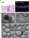
Expanded human airway epithelial cells repopulate a tracheal matrix ex vivo. (A) Hematoxylin and eosin (H&E) staining of rat tracheal xenografts demonstrated a denuded epithelium before human cell engraftment (left; scale bar, 100 μm). Immunofluorescence staining for p63 (green), cytokeratin 5 (CK5; red), and 4′,6-diamidino-2-phenylindole (DAPI; blue) 5 days after seeding of 1 × 107 cells/ml (right; scale bar, 20 μm). (B) Scanning electron microscopy of the xenograft in cross-section 5 days after seeding at 1 × 107 cells/ml (top left; scale bar, 250 μm), the luminal surface shown top down (top right; scale bar, 50 μm), and a higher magnification view of a region with cobblestone epithelial appearance (bottom; scale bar, 1 μm).
Figure 8.
Figure 8.
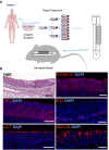
3T3+Y-expanded human airway basal cells contribute to the regeneration in a tracheal xenograft model. (A) Schematic representation of epithelial preparation and xenograft implantation. Human airway mucosal biopsies were expanded in 3T3+Y, engrafted for 6 hours, and implanted subcutaneously in NOD scid gamma mice for 5 weeks. (B) Hematoxylin and eosin (H&E) staining of the restored epithelial layer found throughout the graft (top left). STEM121, a human-specific antibody, revealed regenerated epithelium that was derived from transplanted cells (top right). Immunofluorescence staining for the basal cell markers cytokeratin 5 (CK5) and p63 (middle row), the ciliated cell marker acetylated tubulin (ACT; bottom left), and the goblet cell marker MUC5B (bottom right) revealed a normal organization in regenerated epithelium. All immunofluorescence was costained with 4′,6-diamidino-2-phenylindole (DAPI; blue). Scale bars, 50 μm.

Comment in

References

    1. Kotloff RM, Thabut G. Lung transplantation. Am J Respir Crit Care Med. 2011;184:159–171. - PubMed
    1. Ren X, Ott HC. On the road to bioartificial organs. Pflugers Arch. 2014;466:1847–1857. - PubMed
    1. Macchiarini P, Jungebluth P, Go T, Asnaghi MA, Rees LE, Cogan TA, Dodson A, Martorell J, Bellini S, Parnigotto PP, et al. Clinical transplantation of a tissue-engineered airway. Lancet. 2008;372:2023–2030. - PubMed
    1. Elliott MJ, De Coppi P, Speggiorin S, Roebuck D, Butler CR, Samuel E, Crowley C, McLaren C, Fierens A, Vondrys D, et al. Stem-cell-based, tissue engineered tracheal replacement in a child: a 2-year follow-up study. Lancet. 2012;380:994–1000. - PMC - PubMed
    1. Delaere P, Vranckx J, Verleden G, De Leyn P, Van Raemdonck D Leuven Tracheal Transplant Group. Tracheal allotransplantation after withdrawal of immunosuppressive therapy. N Engl J Med. 2010;362:138–145. - PubMed

Publication types

MeSH terms