

Can Spectral CT Imaging Improve the Differentiation between Malignant and Benign Solitary Pulmonary Nodules?
- PMID: 26840459
- PMCID: PMC4739615
- DOI: 10.1371/journal.pone.0147537
Can Spectral CT Imaging Improve the Differentiation between Malignant and Benign Solitary Pulmonary Nodules?
Abstract
Purpose: To quantitatively assess the value of dual-energy CT (DECT) in differentiating malignancy and benignity of solitary pulmonary nodules.
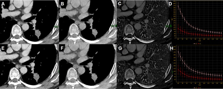
Materials and methods: Sixty-three patients with solitary pulmonary nodules detected by CT plain scan underwent contrast enhanced CT scans in arterial phase (AP) and venous phase (VP) with spectral imaging mode for tumor type differentiation. The Gemstone Spectral Imaging (GSI) viewer was used for image display and data analysis. Region of interest was placed on the relatively homogeneous area of the nodule to measure iodine concentration (IC) on iodine-based material decomposition images and CT numbers on monochromatic image sets to generate spectral HU curve. Normalized IC (NIC), slope of the spectral HU curve (λHU) and net CT number enhancement on 70keV images were calculated. The two-sample t-test was used to compare quantitative parameters. Receiver operating characteristic curves were generated to calculate sensitivity and specificity.
Results: There were 63 nodules, with 37 malignant nodules (59%) and 26 benign nodules (41%). NIC, λHU and net CT number enhancement on 70keV images for malignant nodules were all greater than those of benign nodules. NIC and λHU had intermediate to high performances to differentiate malignant nodules from benign ones with the areas under curve of 0.89 and 0.86 respectively in AP, 0.96 and 0.89 respectively in VP. Using 0.30 as a threshold value for NIC in VP, one could obtain sensitivity of 93.8% and specificity of 85.7% for differentiating malignant from benign solitary pulmonary nodules. These values were statistically higher than the corresponding values of 74.2% and 53.8% obtained with the conventional CT number enhancement.
Conclusions: DECT imaging with GSI mode provides more promising value in quantitative way for distinguishing malignant nodules from benign ones than CT enhancement numbers.
Conflict of interest statement
Figures


References
-
- Winer-Muram HT. The solitary pulmonary nodule. Radiology. 2006;239(1):34–49. - PubMed
-
- Swensen SJ, Yamashita K, McCollough CH, Viggiano RW, Midthun DE, Patz EF Jr, et al. Lung nodules: dual-kilovolt peak analysis with CT—multicenter study. Radiology. 2000;214(1):81–85. - PubMed
-
- Swensen SJ, Viggiano RW, Midthun DE, Müller NL, Sherrick A, Yamashita K, et al. Lung nodule enhancement at CT: multicenter study. Radiology. 2000;214(1): 73–80. - PubMed
Publication types
MeSH terms
LinkOut - more resources
Full Text Sources
Other Literature Sources
Medical
