

Green Fluorescence of Cytaeis Hydroids Living in Association with Nassarius Gastropods in the Red Sea
- PMID: 26840497
- PMCID: PMC4739711
- DOI: 10.1371/journal.pone.0146861
Green Fluorescence of Cytaeis Hydroids Living in Association with Nassarius Gastropods in the Red Sea
Abstract
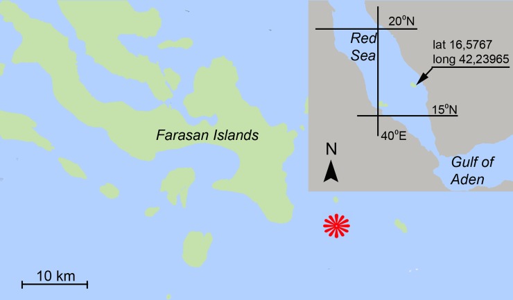
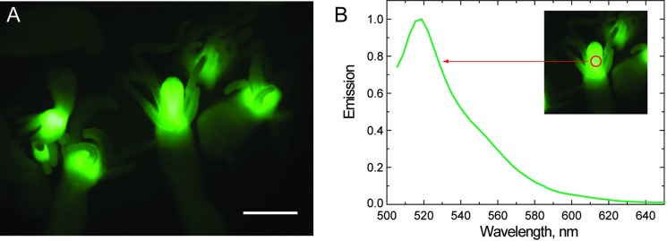
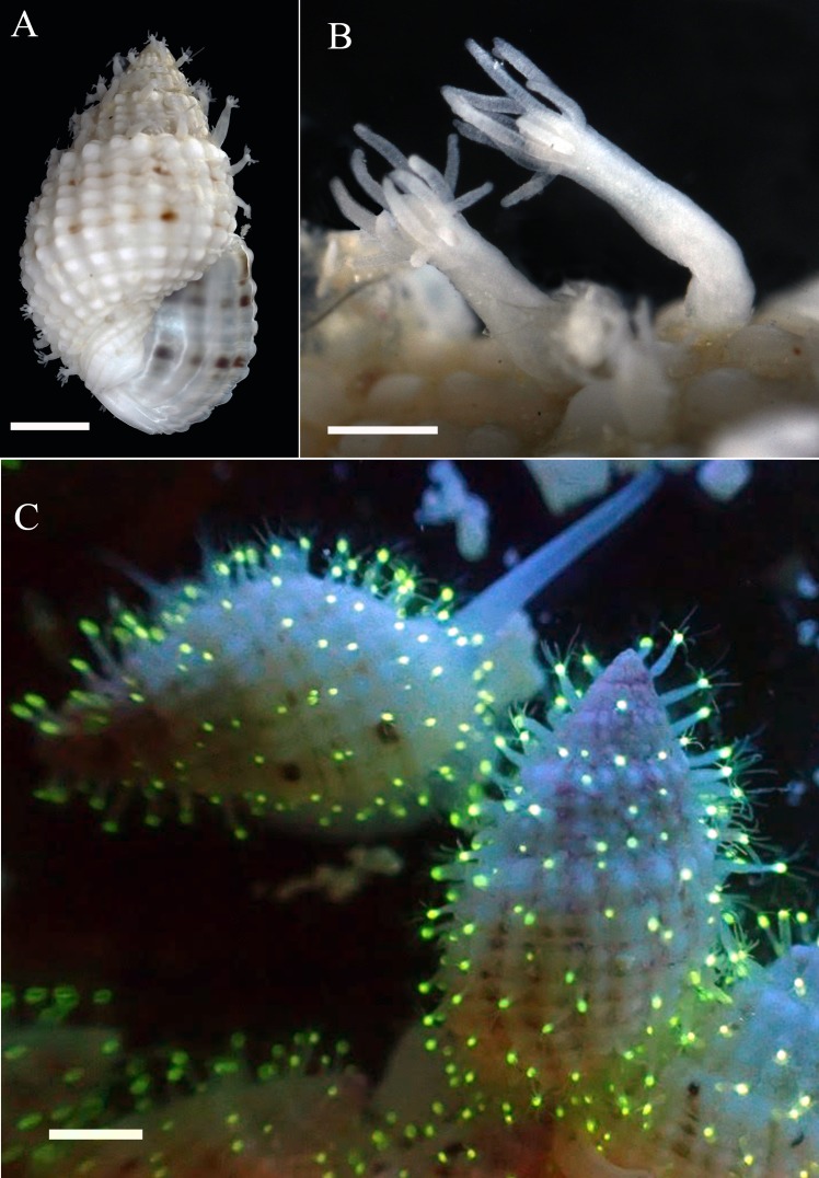
Green Fluorescent Proteins (GFPs) have been reported from a wide diversity of medusae, but only a few observations of green fluorescence have been reported for hydroid colonies. In this study, we report on fluorescence displayed by hydroid polyps of the genus Cytaeis Eschscholtz, 1829 (Hydrozoa: Anthoathecata: Filifera) found at night time in the southern Red Sea (Saudi Arabia) living on shells of the gastropod Nassarius margaritifer (Dunker, 1847) (Neogastropoda: Buccinoidea: Nassariidae). We examined the fluorescence of these polyps and compare with previously reported data. Intensive green fluorescence with a spectral peak at 518 nm was detected in the hypostome of the Cytaeis polyps, unlike in previous reports that reported fluorescence either in the basal parts of polyps or in other locations on hydroid colonies. These results suggest that fluorescence may be widespread not only in medusae, but also in polyps, and also suggests that the patterns of fluorescence localization can vary in closely related species. The fluorescence of polyps may be potentially useful for field identification of cryptic species and study of geographical distributions of such hydroids and their hosts.
Conflict of interest statement
Figures
Similar articles
-
Hydroids (Cnidaria, Hydrozoa) from Mauritanian Coral Mounds.Zootaxa. 2020 Nov 16;4878(3):zootaxa.4878.3.2. doi: 10.11646/zootaxa.4878.3.2. Zootaxa. 2020. PMID: 33311142
-
Diversity and life-cycle analysis of Pacific Ocean zooplankton by videomicroscopy and DNA barcoding: Hydrozoa.PLoS One. 2019 Oct 25;14(10):e0218848. doi: 10.1371/journal.pone.0218848. eCollection 2019. PLoS One. 2019. PMID: 31652271 Free PMC article.
-
The polyps of Oceania armata identified by DNA barcoding (Cnidaria, Hydrozoa).Zootaxa. 2016 Oct 18;4175(6):539-555. doi: 10.11646/zootaxa.4175.6.3. Zootaxa. 2016. PMID: 27811739
-
Molecular phylogenetics of the siphonophora (Cnidaria), with implications for the evolution of functional specialization.Syst Biol. 2005 Dec;54(6):916-35. doi: 10.1080/10635150500354837. Syst Biol. 2005. PMID: 16338764
-
Biology and ecology of the hydrocoral millepora on coral reefs.Adv Mar Biol. 2006;50:1-55. doi: 10.1016/S0065-2881(05)50001-4. Adv Mar Biol. 2006. PMID: 16782450 Review.
References
-
- Shimomura O, Johnson FH, Saiga Y. Extraction, purification and properties of aequorin, a bioluminescent protein from the luminous hydromedusan, Aequorea. J Cell Comp Physiol. 1962;59:223–39. - PubMed
-
- Anderson JM, Cormier MJ. Lumisomes, the cellular site of bioluminescence in coelenterates. J Biol Chem. 1973;248:2937–2943. - PubMed
-
- Morin JG, Reynolds GT. The cellular origin of bioluminescence in the colonial hydroid Obelia. Biol Bull. 1974;147:397–410. - PubMed
Publication types
MeSH terms
Substances
LinkOut - more resources
Full Text Sources
Other Literature Sources
Molecular Biology Databases
