Review
. 2016 Jul;28(27):5509-24.
doi: 10.1002/adma.201504733. Epub 2016 Feb 3.

DNA Origami: Folded DNA-Nanodevices That Can Direct and Interpret Cell Behavior

Affiliations
Review

DNA Origami: Folded DNA-Nanodevices That Can Direct and Interpret Cell Behavior

Cathal J Kearney et al. Adv Mater. 2016 Jul.

Abstract

DNA origami is a DNA-based nanotechnology that utilizes programmed combinations of short complementary oligonucleotides to fold a large single strand of DNA into precise 2D and 3D shapes. The exquisite nanoscale shape control of this inherently biocompatible material is combined with the potential to spatially address the origami structures with diverse cargoes including drugs, antibodies, nucleic acid sequences, small molecules, and inorganic particles. This programmable flexibility enables the fabrication of precise nanoscale devices that have already shown great potential for biomedical applications such as: drug delivery, biosensing, and synthetic nanopore formation. Here, the advances in the DNA-origami field since its inception several years ago are reviewed with a focus on how these DNA-nanodevices can be designed to interact with cells to direct or probe their behavior.

Keywords: DNA origami; biosensing; drug delivery; nanoparticles; nanopores.


Figures

Figure 1
Figure 1
The original DNA origami design technique and shapes. (a) Rothemund[6] used staple oligonucleotides (colored strands) to raster a single continuous scaffold strand (black line) into defined stapes. (b) DNA helix representation of the staples (colored strands) and scaffold (black strands) for the origami technique. (c) Designs for (rows 1 and 2) and AFM images of fabricated DNA origami shapes. Reproduced from [6].
Figure 2
Figure 2
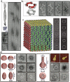
The original 3-D origami folding designs, schematics and sample structures. (a) folding schematic for honeycomb arranged DNA origami lattices[16]. (b) Design and TEM imaging of honeycomb lattice shapes including a box, square nut, railed bridge, slotted cross and stracked cross (scale bar = 20nm) [16]. (c) Folding schematic for square lattice structures [17]. (d) 3- and 6-layer DNA origami cuboid designs and TEM imaging (scale bar 20nm) [17]. Reprinted with permission from[16] and [17] was reprinted with permission © 2009 American Chemical Society.
Figure 3
Figure 3
Twisted, curved and folded DNA origami sheets. (a) DNA nanoribbon based on crossovers every 11 bp/turn of DNA (scale bar 20nm, Lower Left Images; 50nm ribbon image) [18]. (b) Six-tooth gear design developed by carefully bend DNA tubes in the plane (scale bar 20nm) [18]. (c) Hollow DNA origami box design (42 × 36 × 36 nm3), developed by folding a 2-D sheet and stapling along its edges [19]. (D) Strategies for sophisticated in- and out-of-plane curving can lead to intricate DNA origami shapes such as hemispheres, spheres, ellipsoids and nanoflasks (scale bar 50nm) [20]. Reproduced from [18-20].
Figure 4
Figure 4
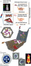
DNA origami strategies for drug delivery. (a) Doxorubicin can be directly intercalated in DNA-origami shapes and shows enhanced localization over controls to tumor regions following injection due to its carefully controlled size and shape that lead to an enhanced permeability and retention (EPR) effect. Once localized at the tumor, it undergoes digestion at the higher pH and releases the active drug. Reprinted with permission from [26]. © 2014 American Chemical Society. (b) Schematic of a DNA nanorobot. The blue and orange tethers on the barrel ends form a lock that is unlocked by aptamers (red globular proteins in the schematic). Once opened, the cargo (pink, which is tethered to the nanorobot via yellow strands) is revealed to the target cell (Reproduced with permission from [37]). (c) A virus-inspired approach to enhancing the circulation time and stability of origami. A DNA NanoOctahedron is encapsulated with a lipid bilayer membrane and when injected in mice shows excellent biodistribution and little bladder accumulations, indicative of it avoiding renal clearance. Reprinted with permission from[57]. © 2014 American Chemical Society.
Figure 5
Figure 5
DNA-origami based synthetic lipid membrane channels. (a) Schematic overview of a synthetic DNA-origami membrane channel that can be used to mimic lipid membrane channel behavior. By adjusting channel size, physical control over the passage of molecules (e.g., dsDNA) can be controlled. Enhanced control can be achieved by extending staple tethers into the channel that can trap ssDNA as it passes. Reprinted with permission from [60]. © 2013 American Chemical Society. (b) Schematic of a DNA origami based synthetic nanopore (Left) and eleven of these synthetic nanopores inserted in a lipid membrane vesicle Reproduced with permission [61].
Figure 6
Figure 6
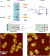
Biosensing DNA-origami logic board for miRNA detection [65]. (a) When the correct miRNA staple strands are detected by the computation section of the board, biotinylated oliognucleotides are unzipped by these miRNA and diffuse to bind to the output section of the board. Following binding to the output strands, streptavidin is introduced and (b) the origami chips read by AFM. Reprinted with permission from [65]. © 2014 American Chemical Society.

Similar articles

Cited by

References

    1. Watson JD, Crick FH. Nature. 1953;171:737. - PubMed
    1. Pray L. Nature Education. 2008;1:100.
    1. Holliday R. Genetics Research. 1964;5:282.
    1. Seeman NC. Journal of theoretical biology. 1982;99:237. - PubMed
    1. Winfree E, Liu F, Wenzler LA, Seeman NC. Nature. 1998;394:539. - PubMed