. 2016 Feb 3;11(2):e0147951.
doi: 10.1371/journal.pone.0147951. eCollection 2016.

The Role of T Cell Costimulation via DNAM-1 in Kidney Transplantation

Affiliations

The Role of T Cell Costimulation via DNAM-1 in Kidney Transplantation

Anna K Kraus et al. PLoS One. .

Abstract

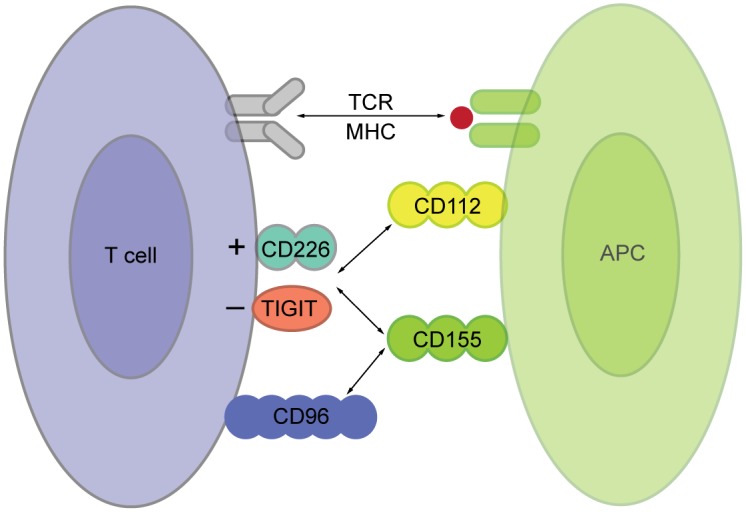
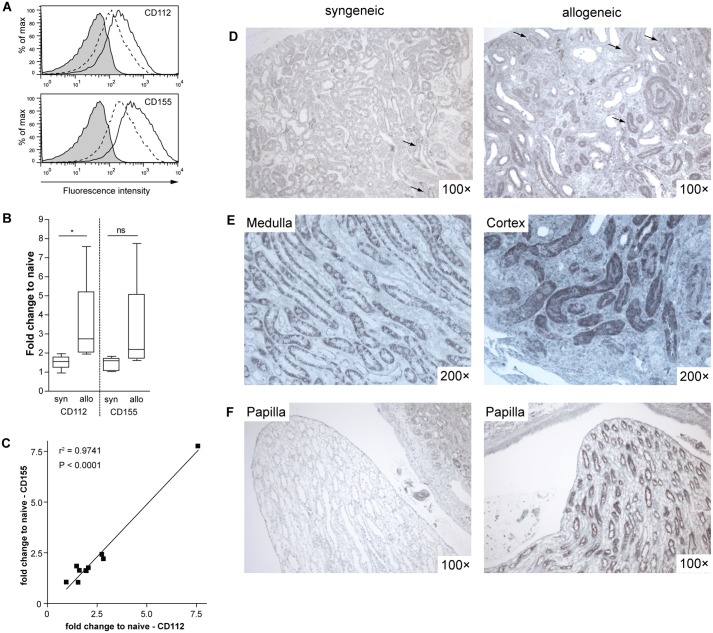
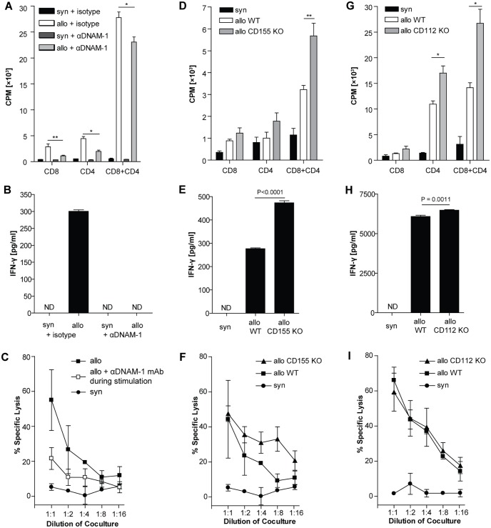
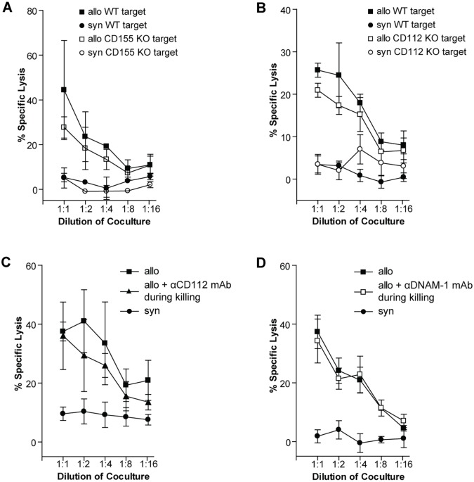
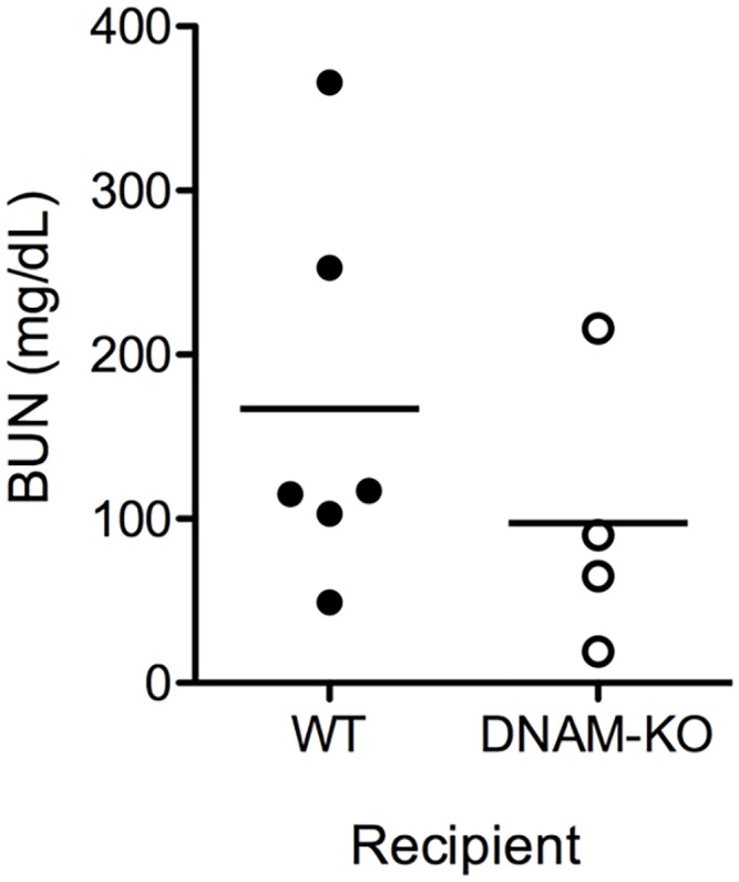
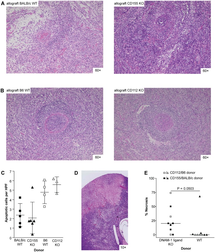
DNAX accessory protein-1 (DNAM-1, CD226) is a co-stimulatory and adhesion molecule expressed mainly by natural killer cells and T cells. DNAM-1 and its two ligands CD112 and CD155 are important in graft-versus-host disease, but their role in solid organ transplantation is largely unknown. We investigated the relevance of this pathway in a mouse kidney transplantation model. CD112 and CD155 are constitutively expressed on renal tubular cells and strongly upregulated in acutely rejected renal allografts. In vitro DNAM-1 blockade during allogeneic priming reduced the allospecific T cell response but not the allospecific cytotoxicity against renal tubular epithelial cells. Accordingly, absence of DNAM-1 in recipient mice or absence of CD112 or CD155 in the kidney allograft did not significantly influence renal function and severity of rejection after transplantation, but led to a higher incidence of infarcts in CD112 and CD155 deficient kidney allografts. Thus, DNAM-1 blockade is not effective in preventing transplant rejection. Despite of being highly expressed, CD112 and CD155 do not appear to play a major immunogenic role in kidney transplantation. Considering the high incidence of renal infarcts in CD112 and CD155 deficient grafts, blocking these molecules might be detrimental.


Conflict of interest statement

Competing Interests: The authors have declared that no competing interests exist.

Figures

Fig 1
Fig 1. DNAM-1 and its two ligands.
Schematic illustration of the DNAM-1 pathway in the interaction between a T cell and an antigen presenting cell (APC). The T cell recognizes its cognate antigen in the context of MHC with its T cell receptor (TCR). For further activation it needs costimulatory signals, which can be delivered via DNAM-1 (CD226) binding to its two ligands CD155 and CD112 expressed on the APC. CD155 and CD112 also bind TIGIT, a co-inhibitory receptor of the Ig-family. CD155 has an additional receptor called CD96.
Fig 2
Fig 2. Renal tubular epithelial cells express CD112 and CD155.
(A) Primary B6-derived rTECs were left untreated or stimulated with IFN-β and -γ (100 U/ml each) for 48 hours. Surface expression of CD155 and CD112 was analyzed by FACS. Shaded: isotype control, dotted line: unstimulated, solid line: stimulated. A representative result of 4 independent experiments is shown. (B-F) Renal allografts from B6 to fully MHC-mismatched CBA recipients were performed. B6 to B6 syngeneic renal grafts were performed as control. In each group we included at least 5 mice. (B) Real time-PCR for CD155 and CD112 was performed on naïve kidneys and renal syn- and allografts. The fold upregulation of CD112 and CD155 compared to naïve renal tissue is depicted. Groups were compared using the Mann-Whitney-test: * P = 0.016, ns = not significant. (C) The expression of CD155 and CD112 in this group of grafts highly correlated (P<0.001). (D-F) Immunohistochemical staining for CD155 was performed. (D) An overview shows the tubular expression of CD155 in both syn- and allografts (arrows, scale bar 200 μm). (E) The expression of CD155 is located mainly in the medulla in syngrafts and is increased in the cortical area in allografts. (F) The papillae of syngrafts show no staining for CD155, whereas in allografts tubuli in the papillae exhibit strong CD155 staining (scale bar 200 μm).
Fig 3
Fig 3. Paradoxical effect of DNAM-1 blockade and CD155/CD112 deficiency.
(A-C) Isolated CBA T cells were stimulated with irradiated B6 splenocytes. CD8 or CD4 T cells were either cultured alone or in combination (ratio 1:2). An anti-DNAM-1 antibody (3B3) was added to the culture at 25 μg/ml. As control an unspecific isotype control antibody was used at the same concentration. (A) Proliferation was measured on day 4 of culture. * P = 0.035, ** P<0.01. (B) IFN-γ was measured in the supernatant from the same CD8+CD4 cultures. (C) Cytotoxicity of CBA splenocytes stimulated in the presence or absence of 3B3 (25 μg/ml) against IFN-stimulated B6 WT rTECs was measured on day 5. (D-F) Isolated B6 T cells were stimulated with irradiated BALB/c WT or CD155 KO splenocytes. CD8 or CD4 T cells were either cultured alone or in combination (ratio 1:2). (D) Proliferation was measured on day 4 of culture. ** P<0.01. (E) IFN-γ was measured in the supernatant from the same CD8+CD4 cultures. (F) Cytotoxicity of the CD8+CD4 culture against IFN-stimulated WT BALB/c rTECs was measured on day 5. (G-I) Isolated CBA T cells were stimulated with irradiated B6 WT or CD112 KO splenocytes. CD8 or CD4 T cells were either cultured alone or in combination (ratio 1:2). (G) Proliferation was measured on day 4 of culture. * P = 0.02. (H) IFN-γ was measured in the supernatant from the same CD8+CD4 cocultures. (I) Cytotoxicity of CBA splenocytes stimulated with irradiated WT B6 or CD112 KO splenocytes against IFN-stimulated WT B6 rTECs was measured on day 5. Data points represent mean values of triplicates. All experiments were performed at least 3 times. Representative figures are displayed.
Fig 4
Fig 4. Cytotoxic activity of allospecific T cells against rTECs is not DNAM-1 dependent in vitro.
(A) CBA splenocytes were stimulated with irradiated BALB/c splenocytes. Cytotoxicity against IFN-stimulated WT BALB/c or CD155 KO rTEC targets was measured on day 5 of coculture. (B) CBA splenocytes were stimulated with irradiated B6 splenocytes. Cytotoxicity against IFN-stimulated WT or CD112 KO targets was measured on day 5 of coculture. (C) CBA splenocytes were stimulated with irradiated BALB/c splenocytes. Cytotoxicity against IFN-stimulated CD155 KO rTEC targets in the presence (25 μg/ml) or absence of a blocking anti-CD112 antibody was measured on day 5 of coculture. (D) CBA splenocytes were stimulated with irradiated B6 splenocytes. Cytotoxicity against IFN-stimulated WT targets was measured on d 5 of coculture in the presence or absence of a blocking anti-DNAM-1 antibody (50 μg/ml). Data points represent mean values of triplicates. All experiments were performed at least 3 times. Representative figures are displayed.
Fig 5
Fig 5. Renal allograft function in DNAM-1 deficient recipients.
Renal transplantation was performed in two steps. First, fully MHC-mismatched CBA kidney allografts were transplanted into WT BALB/c or DNAM-1 deficient mice. The second kidney was removed 7 days after surgery and 3 days later renal graft function was estimated by measurement of serum creatinine und urea. Mean BUN WT 167.2 ± 48.4 mg/dl vs. DNAM1-KO 97.5 ± 42.1 mg/dl, P = 0.36. One mouse in the DNAM1-KO group and 2 mice in the WT group had to be excluded because of surgical complications. Thus, in the final analysis 6 mice were included in the WT group and 4 in the DNAM1-KO group.
Fig 6
Fig 6. Similar rejection but higher rate of infarcts in renal allografts from CD155 or CD112 KO donors.
Renal allografts were performed in a non-life supporting manner. All allografts were harvested on day 21. Strain combinations were fully MHC-mismatched: (A) BALB/c WT (n = 5) or CD155 KO (n = 5) into B6 and (B) B6 WT (n = 5) or CD112 KO (n = 3) into CBA. Representative H & E stainings are shown. All allografts displayed severe interstitial infiltrates as well as tubulitis in more than 50% of the graft. Furthermore, arteritis was detected in all grafts classifying them to Banff grade II or III. The chosen pictures are taken from allografts with the following Banff grades: (A) BALB/c to B6: IIA; CD155 KO to B6: IIB; (B) B6 to CBA: IIB; CD112 to CBA: III (scale bar 200 μm). (C) Apoptotic cells in renal allografts were detected by immunohistochemical staining for ssDNA. (D) Representative picture of a necrotic area in an H & E slide of a CD155 KO renal allograft (scale bar 1200 μm). (E) The area of necrotic tissue in H & E stained slides from renal allografts was detected by scanning them at a resolution of 0.23 μm. Quantification was performed using NDPView software. Groups were compared using the Mann-Whitney-test.

Similar articles

Cited by

References

    1. Kinnear G, Jones ND, Wood KJ. Costimulation blockade: current perspectives and implications for therapy. Transplantation. 2013;95(4):527–35. 10.1097/TP.0b013e31826d4672 - DOI - PMC - PubMed
    1. Kraus AK, Cippa PE, Gaspert A, Chen J, Edenhofer I, Wuthrich RP, et al. Absence of donor CD40 protects renal allograft epithelium and preserves renal function. Transplant international: official journal of the European Society for Organ Transplantation. 2013;26(5):535–44. - PubMed
    1. Pilat N, Sayegh MH, Wekerle T. Costimulatory pathways in transplantation. Semin Immunol. 2011;23(4):293–303. 10.1016/j.smim.2011.04.002 - DOI - PMC - PubMed
    1. Shibuya A, Campbell D, Hannum C, Yssel H, Franz-Bacon K, McClanahan T, et al. DNAM-1, a novel adhesion molecule involved in the cytolytic function of T lymphocytes. Immunity. 1996;4(6):573–81. - PubMed
    1. Seth S, Georgoudaki AM, Chambers BJ, Qiu Q, Kremmer E, Maier MK, et al. Heterogeneous expression of the adhesion receptor CD226 on murine NK and T cells and its function in NK-mediated killing of immature dendritic cells. J Leukoc Biol. 2009;86(1):91–101. 10.1189/jlb.1208745 - DOI - PubMed

Publication types

MeSH terms

Substances