

Analysis of fracture healing in osteopenic bone caused by disuse: experimental study
- PMID: 26840708
- PMCID: PMC4763822
- DOI: 10.1590/1414-431X20155076
Analysis of fracture healing in osteopenic bone caused by disuse: experimental study
Abstract
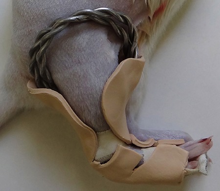
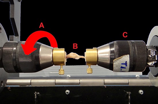
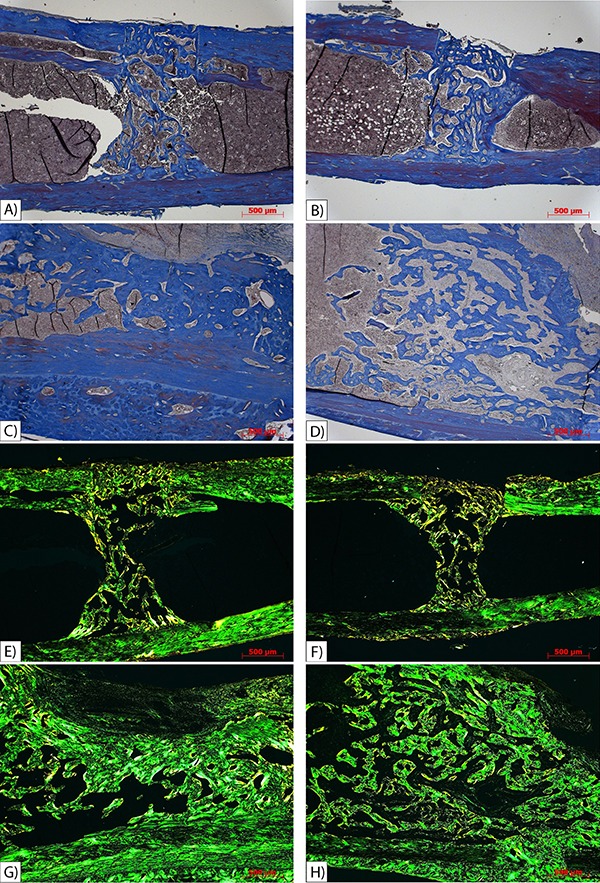
Osteoporosis has become a serious global public health issue. Hence, osteoporotic fracture healing has been investigated in several previous studies because there is still controversy over the effect osteoporosis has on the healing process. The current study aimed to analyze two different periods of bone healing in normal and osteopenic rats. Sixty, 7-week-old female Wistar rats were randomly divided into four groups: unrestricted and immobilized for 2 weeks after osteotomy (OU2), suspended and immobilized for 2 weeks after osteotomy (OS2), unrestricted and immobilized for 6 weeks after osteotomy (OU6), and suspended and immobilized for 6 weeks after osteotomy (OS6). Osteotomy was performed in the middle third of the right tibia 21 days after tail suspension, when the osteopenic condition was already set. The fractured limb was then immobilized by orthosis. Tibias were collected 2 and 6 weeks after osteotomy, and were analyzed by bone densitometry, mechanical testing, and histomorphometry. Bone mineral density values from bony calluses were significantly lower in the 2-week post-osteotomy groups compared with the 6-week post-osteotomy groups (multivariate general linear model analysis, P<0.000). Similarly, the mechanical properties showed that animals had stronger bones 6 weeks after osteotomy compared with 2 weeks after osteotomy (multivariate general linear model analysis, P<0.000). Histomorphometry indicated gradual bone healing. Results showed that osteopenia did not influence the bone healing process, and that time was an independent determinant factor regardless of whether the fracture was osteopenic. This suggests that the body is able to compensate for the negative effects of suspension.
Figures
References
-
- Szejnfeld VL, Jennings F, Castro CHM, Pinheiro MM, Lopes AC. Conhecimento dos médicos clínicos do Brasil sobre as estratégias de prevenção e tratamento da osteoporose. Rev Bras Reumatol. 2015;47:251–257. doi: 10.1590/S0482-50042007000400003. - DOI
MeSH terms
Substances
LinkOut - more resources
Full Text Sources
Other Literature Sources
Medical
