

Identification of new FGF1 binding partners-Implications for its intracellular function
- PMID: 26840910
- PMCID: PMC4832500
- DOI: 10.1002/iub.1480
Identification of new FGF1 binding partners-Implications for its intracellular function
Abstract
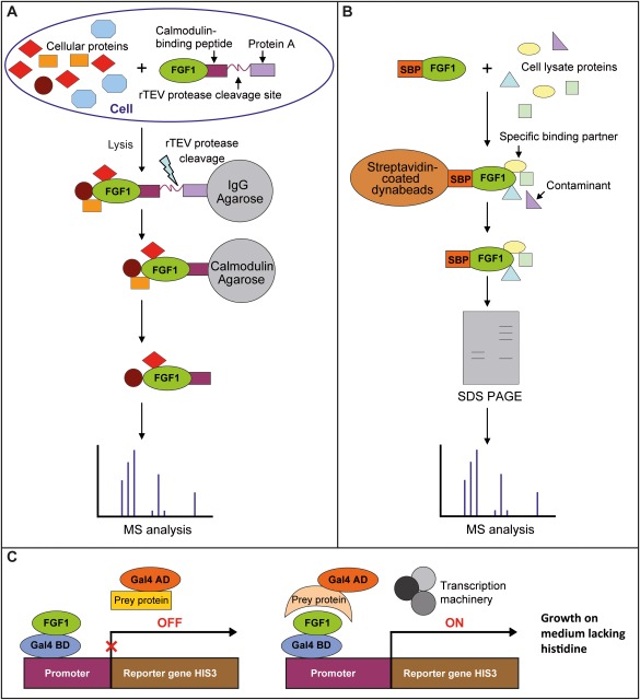
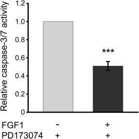
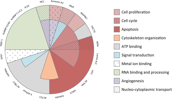
Besides its classical mode of action through activation of specific receptors at the cell surface, fibroblast growth factor 1 (FGF1) can also cross the cellular membrane and translocate into the cytosol and further to the nucleus. The mechanism of this translocation is described partially, but the role of FGF1 inside the cell remains unknown. The aim of our work was to identify novel binding partners of FGF1 to predict its intracellular functions. We combined three methods of identification of such partners based on different principles: yeast two-hybrid screen and mass spectrometry (MS) analysis of complexes obtained by Tandem Affinity Purification (TAP) or by co-precipitation from cell lysate using recombinant FGF1. Altogether, we identified twenty novel intracellular proteins interacting with FGF1. For selected proteins, their direct interaction with FGF1 was confirmed by pull-down assays and SPR measurements. Interestingly, half of the proteins found are involved in processes related to cell viability, such as apoptosis, cell proliferation, and cell cycle regulation. Thus, our study indicates that the role of intracellular FGF1 is to protect the cell against stress conditions by providing an additional signal for cell survival, independently of receptor-activated signaling cascades.
Keywords: FGF1; Tandem Affinity Purification; apoptosis; binding partners; cell survival; intracellular function; translocation.
© 2016 The Authors IUBMB Life published by Wiley Periodicals, Inc. on behalf of International Union of Biochemistry and Molecular Biology.
Figures
Similar articles
-
Translocation of Exogenous FGF1 and FGF2 Protects the Cell against Apoptosis Independently of Receptor Activation.J Mol Biol. 2018 Oct 19;430(21):4087-4101. doi: 10.1016/j.jmb.2018.08.004. Epub 2018 Aug 9. J Mol Biol. 2018. PMID: 30099027
-
Nucleolin regulates phosphorylation and nuclear export of fibroblast growth factor 1 (FGF1).PLoS One. 2014 Mar 4;9(3):e90687. doi: 10.1371/journal.pone.0090687. eCollection 2014. PLoS One. 2014. PMID: 24595027 Free PMC article.
-
Size limitation in translocation of fibroblast growth factor 1 fusion proteins across the endosomal membrane.Biochemistry. 2009 Aug 4;48(30):7209-18. doi: 10.1021/bi9007353. Biochemistry. 2009. PMID: 19558187
-
Intracellular partners of fibroblast growth factors 1 and 2 - implications for functions.Cytokine Growth Factor Rev. 2021 Feb;57:93-111. doi: 10.1016/j.cytogfr.2020.05.004. Epub 2020 May 18. Cytokine Growth Factor Rev. 2021. PMID: 32475760 Review.
-
The non-classical export routes: FGF1 and IL-1alpha point the way.J Cell Sci. 2003 Dec 15;116(Pt 24):4871-81. doi: 10.1242/jcs.00872. J Cell Sci. 2003. PMID: 14625381 Review.
Cited by
-
Proteomic Analysis of Potential Targets for Non-Response to Infliximab in Patients With Ulcerative Colitis.Front Pharmacol. 2022 Jun 13;13:905133. doi: 10.3389/fphar.2022.905133. eCollection 2022. Front Pharmacol. 2022. PMID: 35770079 Free PMC article.
-
Kallikrein-related peptidase 4 induces cancer-associated fibroblast features in prostate-derived stromal cells.Mol Oncol. 2017 Oct;11(10):1307-1329. doi: 10.1002/1878-0261.12075. Epub 2017 Aug 10. Mol Oncol. 2017. PMID: 28510269 Free PMC article.
-
FGF1 induces resistance to chemotherapy in ovarian granulosa tumor cells through regulation of p53 mitochondrial localization.Oncogenesis. 2018 Feb 21;7(2):18. doi: 10.1038/s41389-018-0033-y. Oncogenesis. 2018. PMID: 29467390 Free PMC article.
-
Intracellular FGF1 protects cells from apoptosis through direct interaction with p53.Cell Mol Life Sci. 2023 Oct 2;80(10):311. doi: 10.1007/s00018-023-04964-9. Cell Mol Life Sci. 2023. PMID: 37783936 Free PMC article.
-
New developments in the biology of fibroblast growth factors.WIREs Mech Dis. 2022 Jul;14(4):e1549. doi: 10.1002/wsbm.1549. Epub 2022 Feb 9. WIREs Mech Dis. 2022. PMID: 35142107 Free PMC article. Review.
References
-
- Powers, C. J. , McLeskey, S. W. , and Wellstein, A. (2000) Fibroblast growth factors, their receptors and signaling. Endocr. Relat. Cancer 7, 165–197. - PubMed
-
- Schlessinger, J. (2000) Cell signaling by receptor tyrosine kinases. Cell 103, 211–225. - PubMed
-
- Eswarakumar, V. P. , Lax, I. , and Schlessinger, J. (2005) Cellular signaling by fibroblast growth factor receptors. Cytokine Growth Factor Rev. 16, 139–146. - PubMed
-
- Wiedlocha, A. and Sorensen, V. (2004) Signaling, internalization, and intracellular activity of fibroblast growth factor. Curr. Top. Microbiol. Immunol. 286, 45–79. - PubMed
Publication types
MeSH terms
Substances
LinkOut - more resources
Full Text Sources
Other Literature Sources
Miscellaneous
