

The external globus pallidus: progress and perspectives
- PMID: 26841063
- PMCID: PMC4874844
- DOI: 10.1111/ejn.13196
The external globus pallidus: progress and perspectives
Abstract
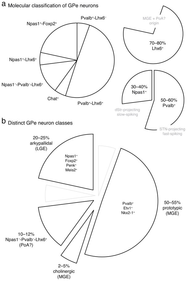
The external globus pallidus (GPe) of the basal ganglia is in a unique and powerful position to influence processing of motor information by virtue of its widespread projections to all basal ganglia nuclei. Despite the clinical importance of the GPe in common motor disorders such as Parkinson's disease, there is only limited information about its cellular composition and organizational principles. In this review, recent advances in the understanding of the diversity in the molecular profile, anatomy, physiology and corresponding behaviour during movement of GPe neurons are described. Importantly, this study attempts to build consensus and highlight commonalities of the cellular classification based on existing but contentious literature. Additionally, an analysis of the literature concerning the intricate reciprocal loops formed between the GPe and major synaptic partners, including both the striatum and the subthalamic nucleus, is provided. In conclusion, the GPe has emerged as a crucial node in the basal ganglia macrocircuit. While subtleties in the cellular makeup and synaptic connection of the GPe create new challenges, modern research tools have shown promise in untangling such complexity, and will provide better understanding of the roles of the GPe in encoding movements and their associated pathologies.
Keywords: Npas1; Parkinson's disease; arkypallidal neurons; parvalbumin; prototypic neurons.
© 2016 Federation of European Neuroscience Societies and John Wiley & Sons Ltd.
Figures
Similar articles
-
Parvalbumin+ and Npas1+ Pallidal Neurons Have Distinct Circuit Topology and Function.J Neurosci. 2020 Oct 7;40(41):7855-7876. doi: 10.1523/JNEUROSCI.0361-20.2020. Epub 2020 Aug 31. J Neurosci. 2020. PMID: 32868462 Free PMC article.
-
Sensory processing in external globus pallidus neurons.Cell Rep. 2023 Jan 31;42(1):111952. doi: 10.1016/j.celrep.2022.111952. Epub 2023 Jan 4. Cell Rep. 2023. PMID: 36640317
-
Dysregulation of external globus pallidus-subthalamic nucleus network dynamics in parkinsonian mice during cortical slow-wave activity and activation.J Physiol. 2020 May;598(10):1897-1927. doi: 10.1113/JP279232. Epub 2020 Apr 23. J Physiol. 2020. PMID: 32112413 Free PMC article.
-
Connectivity and Functionality of the Globus Pallidus Externa Under Normal Conditions and Parkinson's Disease.Front Neural Circuits. 2021 Mar 2;15:645287. doi: 10.3389/fncir.2021.645287. eCollection 2021. Front Neural Circuits. 2021. PMID: 33737869 Free PMC article.
-
Move to the rhythm: oscillations in the subthalamic nucleus-external globus pallidus network.Trends Neurosci. 2002 Oct;25(10):525-31. doi: 10.1016/s0166-2236(02)02235-x. Trends Neurosci. 2002. PMID: 12220881 Review.
Cited by
-
Selective encoding of reward predictions and prediction errors by globus pallidus subpopulations.Curr Biol. 2023 Oct 9;33(19):4124-4135.e5. doi: 10.1016/j.cub.2023.08.042. Epub 2023 Sep 12. Curr Biol. 2023. PMID: 37703876 Free PMC article.
-
Blunted mGluR Activation Disinhibits Striatopallidal Transmission in Parkinsonian Mice.Cell Rep. 2016 Nov 22;17(9):2431-2444. doi: 10.1016/j.celrep.2016.10.087. Cell Rep. 2016. PMID: 27880915 Free PMC article.
-
Striosomes Target Nigral Dopamine-Containing Neurons via Direct-D1 and Indirect-D2 Pathways Paralleling Classic Direct-Indirect Basal Ganglia Systems.bioRxiv [Preprint]. 2024 Jul 23:2024.06.01.596922. doi: 10.1101/2024.06.01.596922. bioRxiv. 2024. PMID: 38915684 Free PMC article. Preprint.
-
Spontaneous Activity of the Local GABAergic Synaptic Network Causes Irregular Neuronal Firing in the External Globus Pallidus.J Neurosci. 2023 Feb 22;43(8):1281-1297. doi: 10.1523/JNEUROSCI.1969-22.2023. Epub 2023 Jan 9. J Neurosci. 2023. PMID: 36623877 Free PMC article.
-
Lesions causing post-stroke spasticity localize to a common brain network.Front Aging Neurosci. 2022 Oct 25;14:1011812. doi: 10.3389/fnagi.2022.1011812. eCollection 2022. Front Aging Neurosci. 2022. PMID: 36389077 Free PMC article.
References
-
- Adler A, Joshua M, Rivlin-Etzion M, Mitelman R, Marmor O, Prut Y, Bergman H. Neurons in both pallidal segments change their firing properties similarly prior to closure of the eyes. J Neurophysiol. 2010;103:346–359. - PubMed
-
- Agari T, Yasuhara T, Matsui T, Kuramoto S, Kondo A, Miyoshi Y, Shingo T, Borlongan CV, Date I. Intrapallidal metabotropic glutamate receptor activation in a rat model of Parkinson’s disease: behavioral and histological analyses. Brain research. 2008;1203:189–196. - PubMed
Publication types
MeSH terms
Grants and funding
LinkOut - more resources
Full Text Sources
Other Literature Sources
