. 2016 Mar 25;291(13):6796-812.
doi: 10.1074/jbc.M115.684787. Epub 2016 Feb 3.

Sorting Motifs in the Cytoplasmic Tail of the Immunomodulatory E3/49K Protein of Species D Adenoviruses Modulate Cell Surface Expression and Ectodomain Shedding

Affiliations

Sorting Motifs in the Cytoplasmic Tail of the Immunomodulatory E3/49K Protein of Species D Adenoviruses Modulate Cell Surface Expression and Ectodomain Shedding

Mark Windheim et al. J Biol Chem. .

Abstract

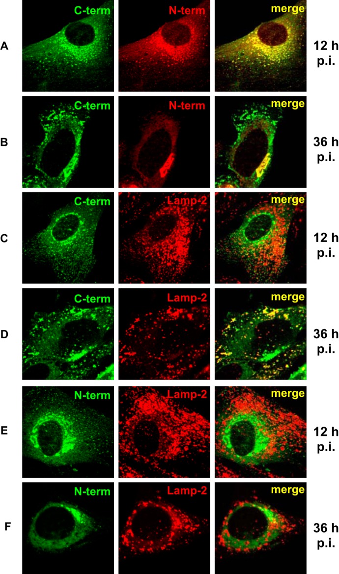
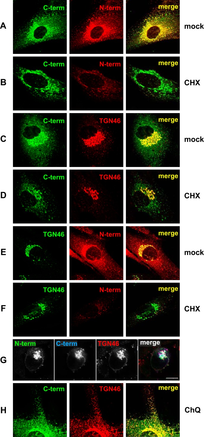
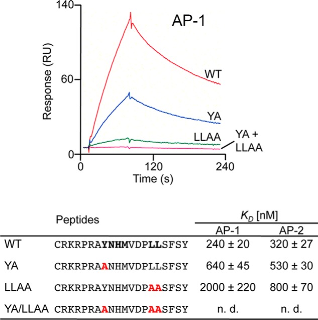
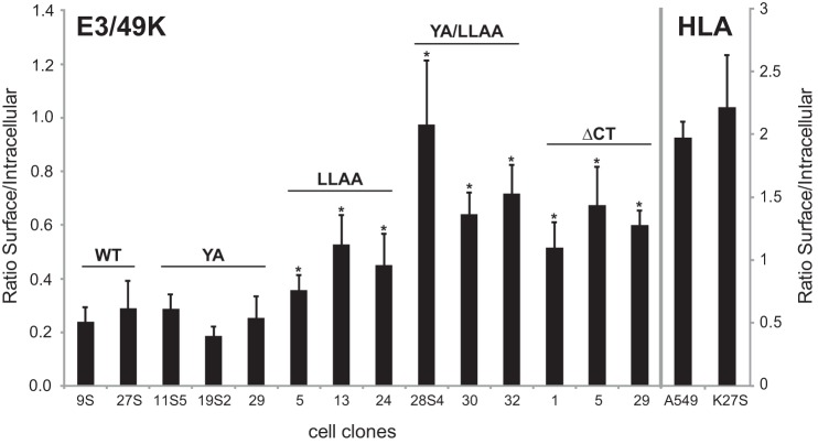
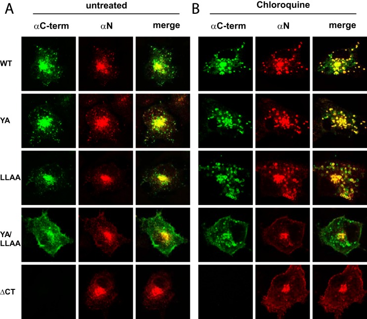
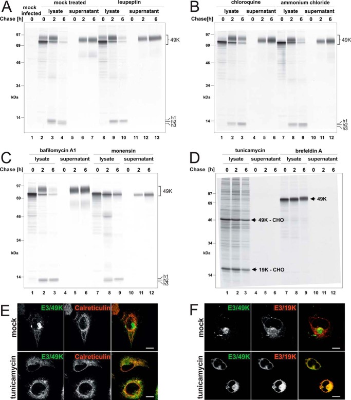
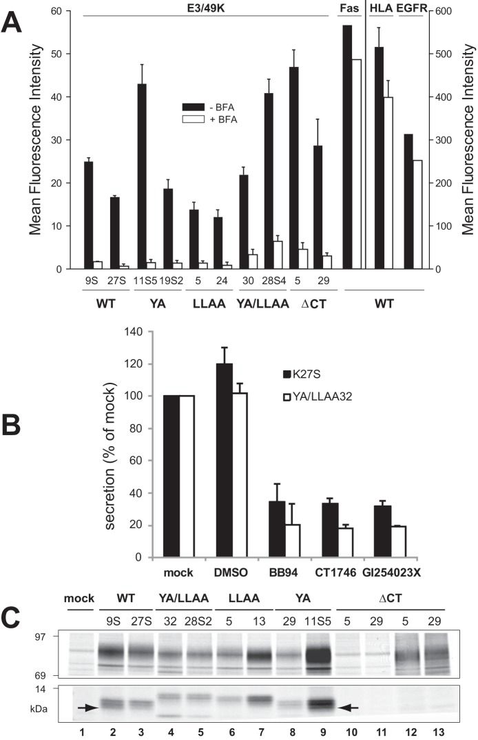
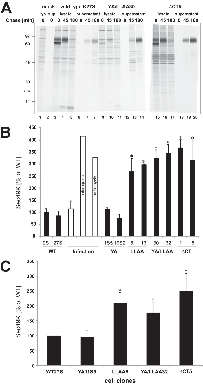
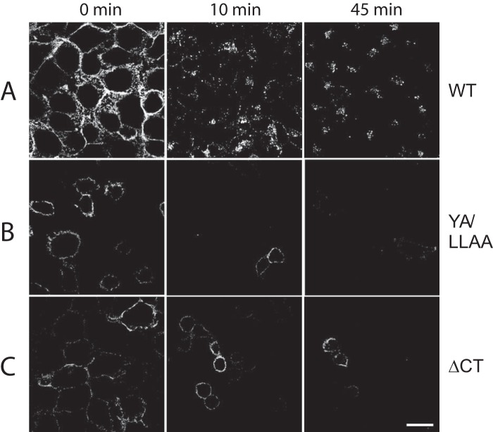
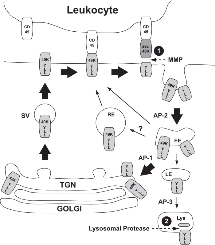
The E3 transcription unit of human species C adenoviruses (Ads) encodes immunomodulatory proteins that mediate direct protection of infected cells. Recently, we described a novel immunomodulatory function for E3/49K, an E3 protein uniquely expressed by species D Ads. E3/49K of Ad19a/Ad64, a serotype that causes epidemic keratokonjunctivitis, is synthesized as a highly glycosylated type I transmembrane protein that is subsequently cleaved, resulting in secretion of its large ectodomain (sec49K). sec49K binds to CD45 on leukocytes, impairing activation and functions of natural killer cells and T cells. E3/49K is localized in the Golgi/trans-Golgi network (TGN), in the early endosomes, and on the plasma membrane, yet the cellular compartment where E3/49K is cleaved and the protease involved remained elusive. Here we show that TGN-localized E3/49K comprises both newly synthesized and recycled molecules. Full-length E3/49K was not detected in late endosomes/lysosomes, but the C-terminal fragment accumulated in this compartment at late times of infection. Inhibitor studies showed that cleavage occurs in a post-TGN compartment and that lysosomotropic agents enhance secretion. Interestingly, the cytoplasmic tail of E3/49K contains two potential sorting motifs, YXXΦ (where Φ represents a bulky hydrophobic amino acid) and LL, that are important for binding the clathrin adaptor proteins AP-1 and AP-2in vitro Surprisingly, mutating the LL motif, either alone or together with YXXΦ, did not prevent proteolytic processing but increased cell surface expression and secretion. Upon brefeldin A treatment, cell surface expression was rapidly lost, even for mutants lacking all known endocytosis motifs. Together with immunofluorescence data, we propose a model for intracellular E3/49K transport whereby cleavage takes place on the cell surface by matrix metalloproteases.

Keywords: Adenovirus E3 proteins; CD45; E3/49K; Epidemic Keratoconjunctivitis; Immune evasion; adenovirus; intracellular trafficking; protein sorting; shedding; viral immunology.


Figures

FIGURE 1.
FIGURE 1.
Differential localization of the ectodomain and the cytoplasmic tail of E3/49K in the Golgi/TGN and late endosomes/lysosomes during early and late phases of infection. Primary fibroblasts (SeBu) were infected with Ad19a and processed for confocal laser microscopy at 12 or 36 h postinfection (p.i.), corresponding to the early and late phase of the infection cycle in these primary fibroblasts, respectively. The subcellular localization of E3/49K was analyzed with rabbit antiserum R25050 directed against the C terminus (C-term; A–D) and antiserum R48-7B (anti-49K-N; E and F), followed by FITC-labeled goat anti-rabbit IgG. For detection of the N-terminal part of E3/49K (N-term) the rat mAb 4D1 was employed (A and B, middle). This was compared with the late endosomal/lysosomal marker LAMP-2, using mouse mAb 2D5 (C and F, middle). Subsequent staining was done with donkey anti-rat IgG Texas Red and donkey anti-mouse IgG Rhodamine Red-X, respectively. One typical experiment of at least three is shown.
FIGURE 2.
FIGURE 2.
The steady state localization of E3/49K in the Golgi/TGN comprises newly synthesized uncleaved E3/49K as well as recycled C-terminal fragments that co-localize extensively with TGN46. Primary fibroblasts (SeBu) infected with Ad19a for 12 h were treated for 6 h with 50 μg/ml CHX. Subsequently, cells were processed for confocal laser microscopy by staining E3/49K with anti-49Kct (A–D) and rat mAb 4D1 against the ectodomain (N-term; A, B, E, and F). Golgi/TGN localization was determined by co-staining for the TGN marker TGN46 using either sheep polyclonal Abs (C, D, G, and H) or rabbit polyclonal Abs (E and F), followed by FITC-labeled donkey anti-rabbit IgG or rhodamine-labeled donkey anti-sheep IgG, anti-rat IgG, and anti-mouse IgG. For the triple labeling experiment (G), A549 cells infected with Ad19a for 9 h were fixed and stained with antibodies against TGN46 and the N and C termini of E3/49K. Secondary Abs were Alexa Fluor 488-labeled goat anti-rat IgG, Alexa Fluor 633-labeled goat anti-rabbit IgG, and Alexa Fluor 594-labeled donkey anti-sheep IgG. Images are single optical sections. Each channel was imaged separately under single laser illumination to prevent cross-over and overlaid using Leica confocal software. Scale bar, 10 μm. H, primary fibroblasts infected with Ad19a for 12 h were treated for 3 h with 100 μm chloroquine (CHQ) prior to staining with antibodies against TGN46 and anti-49Kct. The figure shows one typical experiment of three (A–F) and two (G and H), respectively.
FIGURE 3.
FIGURE 3.
Efficient binding of clathrin adaptor proteins AP-1 and AP-2 to cytoplasmic tail peptides of E3/49K depends on the presence of the YXXΦ and LL motifs. The bottom panel shows the different cytoplasmic tail peptides used for the surface plasmon resonance spectroscopy studies with putative sorting signals in boldface type. YA is a mutant version with alanine substitution of the YXXΦ motif; LLAA is an altered version with alanine substitution of the LL motif; and YA/LLAA is a peptide with both motifs eliminated. Altered amino acids are shown in red. The equilibrium constants (KD) were determined in at least three independent experiments and are given in nm/liter ± S.D. The top panel shows the response versus time in seconds for the incubation of the different peptides with purified AP-1. n.d., not detectable. RU, resonance units.
FIGURE 4.
FIGURE 4.
Elimination of the LL motif in the cytoplasmic tail significantly enhances relative cell surface expression of E3/49K. A549 cell lines stably expressing WT E3/49K or mutants in which the LL and YXXΦ motif, respectively, in the cytoplasmic tail were mutated to AA (LLAA) and AXXΦ (YA). In YA/LLAA, both putative sorting motifs were mutated, whereas ΔCT lacked the entire cytoplasmic tail except a basic RKR stop-transfer sequence. Expression on the cell surface and internally was quantitatively analyzed by flow cytometry in the absence and presence of the detergent saponin, respectively. By determining the ratio of cell surface versus intracellular MFIs rather than the absolute cell surface values, E3/49K cell surface expression becomes independent of its general synthesis level in each transfectant clone. MFIs for cell surface and intracellular staining were determined in 4–6 independent experiments for 2–5 isolated clones, of which 2–3 are shown. Error bars, S.D. A significant difference between cell lines expressing wild type and mutated E3/49K is indicated by an asterisk (p < 0.05). The difference in the ratios between double mutant YA/LLAA and ΔCT is not statistically significant.
FIGURE 5.
FIGURE 5.
Removal of both putative sorting motifs enhances cell surface display of full-length E3/49K and C-terminal fragments and reduces their accumulation in swollen vesicles during chloroquine treatment. A549 cells were transiently transfected with pSG5 plasmids encoding WT and mutant E3/49K proteins as described under “Experimental Procedures.” Cells were treated with 100 μm chloroquine for 12 h (B) or left untreated (A). After 36 h, cells were processed for confocal laser microscopy. Staining was performed with rat mAb 4D1 directed against the N-terminal ectodomain (αN) and rabbit serum R25050 against the C terminus of E3/49K (αC-term), followed by donkey anti-rabbit IgG FITC and donkey anti-mouse IgG Rhodamine Red-X. The figure shows a representative experiment of three.
FIGURE 6.
FIGURE 6.
Proteolytic processing and secretion of E3/49K in the presence of agents affecting glycosylation, endosomal/lysosomal proteases, the pH of intracellular compartments, or trafficking. Ad19a-infected A549 cells were labeled with [35S]methionine for 30 min (0 h) and chased with medium containing non-radioactive methionine for 2 and 6 h. Subsequently, detergent extracts were prepared (lysate), and E3/49K was immunoprecipitated with anti-49Kct, whereas the supernatant was processed and immunoprecipitated with anti-49K-N as described (32). The different agents indicated at the top were included both in the starvation medium and the following incubations. A, mock-treated, with the same medium changes but without inhibitors (lanes 2–7) and leupeptin (200 μm; lanes 8–13); B, chloroquine (100 μm; lanes 1–6) and ammonium chloride (10 mm; lanes 7–12); C, bafilomycin A1 (100 nm; lanes 1–6) and monensin (10 μm; lanes 7–12); D, tunicamycin (10 μg/ml; lanes 1–6) and brefeldin A (5 μg/ml; lanes 7–12). The high molecular weight E3/49K species (49K) and the C-terminal fragments h1–h3 are denoted on the right. E3/49K-CHO and E3/19K-CHO, the unglycosylated species of E3/49K and E3/19K. E, deglycosylated E3/49K colocalizes with the ER marker calreticulin upon tunicamycin treatment (3 h) in permanently transfected A549 cells expressing codon-optimized E3/49K (A549E3/49K-co15) and in infected cells (data not shown). Calreticulin was detected with a rabbit antiserum (Stressgen). F, A549 cells were infected with Ad19a for 6 h and treated for 3 h with tunicamycin (bottom panels) or were mock-treated (DMSO; top panels). Fixed cells were stained for E3/49K N terminus and the ER-resident Ad19a protein E3/19K using rabbit antiserum R22612 and imaged as in Fig. 2G. Scale bar, 20 μm. All experiments were carried out at least twice.
FIGURE 7.
FIGURE 7.
E3/49K cleavage occurs independent of the putative transport motifs at the cell surface and is mediated by MMPs. A, the mean fluorescence intensity of E3/49K WT or mutant clones (indicated at the bottom) on the cell surface was determined by flow cytometry using mAb 4D1 after mock (black bars) and 4-h BFA treatment (5 μg/ml; white bars). Mean values and the S.D. were calculated from 4–6 independent experiments except for Fas and EGF receptor expression that was measured only once. No BFA-induced cytotoxicity or increased background staining was detectable within this time frame. B, cells expressing WT E3/49K (K27S) or the double mutant YA/LLAA (clone 32) lacking all motifs were incubated with the broad spectrum MMP inhibitors BB-94 and CT1746 and the more selective ADAM10 inhibitor GI254023X in 0.1% DMSO (final concentration) for 15–18 h. Cells were also incubated with medium alone (mock) and 0.1% DMSO as solvent control (DMSO). Supernatants were harvested and centrifuged at 1500 × g. Jurkat T cells (6 × 10⁶) were incubated with 200 μl of supernatant, followed by detection of sec49K binding using FACS analysis. The data show the mean plus S.D. (error bars) of five independent staining experiments and three independently produced supernatants. C, mutation of the transport motifs does not prevent initial cleavage but rather secondary cleavage. Transfected clones indicated at the top expressing E3/49K WT and mutant proteins were metabolically labeled for 2 h. E3/49K was precipitated with anti-49Kct antiserum (lanes 1–11) and anti-49K-N (lanes 12 and 13). The figure shows one of two experiments. Only the relevant sections of the SDS-PAGE with the 80–100- and 10–14-kDa E3/49K species are shown. The arrow marks the C-terminal fragment h3.
FIGURE 8.
FIGURE 8.
Elimination of the LL motif alone or in combination with the YXXΦ motif enhances secretion. A, the processing kinetics of WT and mutant E3/49K proteins was determined by a 30-min pulse with [35S]methionine followed by a 45- and 180-min chase. E3/49K was precipitated from untransfected (mock), WT, and YA/LLAA mutant cell lysates with anti-49Kct (lanes 1–5 and 9–11) and anti-49K-N from ΔCT mutant cell lysates (lanes 15–17) and all supernatants (lanes 6–8, 12–14, and 18–20). B, the proportion of E3/49K secreted was quantitatively determined in a separate pulse (0.5 h)-chase (4 h) experiment followed by immunoprecipitation with anti-49K-N and SDS-PAGE. Bands corresponding to sec49K were quantified by phosphorimaging analysis and related to the radioactivity in E3/49K high molecular weight species after the pulse that was defined as 100%. The maximum amount is secreted when 80% of the total E3/49K labeling is released because the cytoplasmic tail contains 20% of the total [35S]methionine labeling. Mean values were calculated from two independent experiments except for the values obtained upon chloroquine and bafilomycin A1 treatment, which were derived from single experiments. The mean value of WT clone K9S was set as 100%. Error bars, S.D. A significant difference between mutants and WT is indicated by asterisks (p < 0.05). C, enhanced lymphocyte binding activity (sec49K) is observed in supernatants of mutants lacking the LL motif. One clone from WT and each mutant was assessed for the efficiency of secretion as measured using the Jurkat T lymphocyte binding assay. Cells were cultured for 4 days, and the supernatant was harvested and centrifuged. Three independently collected supernatants were analyzed for the presence of sec49K in at least four different experiments as described under “Experimental Procedures.” Error bars, S.D. A significant difference between mutants and WT is indicated by asterisks (p < 0.05).
FIGURE 9.
FIGURE 9.
Surface fluorescence of mutated E3/49K is lost in the absence of any internalization, whereas WT E3/49K is internalized. A549 cells stably expressing either WT, YA/LLAA mutant, or ΔCT truncated E3/49K were surface-labeled on ice with E3/49K N-terminal mAb 4D1. Excess antibody was removed, and then cells were warmed to 37 °C and incubated for the times indicated. Upon fixation, the location of prebound antibody was then revealed by staining with secondary antibody and imaged as in Fig. 2G using the same settings throughout to ensure comparability of image intensity. Scale bar, 25 μm. One representative experiment of at least two is shown.
FIGURE 10.
FIGURE 10.
Model for E3/49K trafficking and proteolytic processing. At steady state, E3/49K is found in the Golgi/TGN, at the plasma membrane, and in EEs. At the TGN, E3/49K seems to be packaged by default into secretory vesicles (SV); however, a diversion to an EE prior to appearing on the cell surface cannot formally be ruled out. Proteolytic cleavage (dotted arrows) releasing the N-terminal ectodomain most likely occurs at the cell surface through “shedding” by MMPs (1). The C-terminal fragment and a considerable fraction of uncleaved E3/49K appear to be endocytosed through interaction of the LL motif in the C-tail with AP-2 (thick arrows). Full-length E3/49K may be sorted in the EEs into a recycling pathway to the plasma membrane, either directly from EEs or via the TGN or the recycling endosome (RE) for another round of cell surface transport possibly involving AP-1. A minor pathway may direct E3/49K or predominantly the cytoplasmic tail fragments to LE and lysosomes (Lys), where further proteolytic processing of h1/2 to h3 takes place (3). For further details and references, see “Results.” The extent of the proposed transport pathway is indicated by the thickness of the arrows.

References

    1. King A. M. Q., Adams M. J., Carstens E. B., and Lefkowitz E. J. (eds) (2012) Virus Taxonomy: Classification and Nomenclature of Viruses: Ninth Report of the International Committee on Taxonomy of Viruses, pp. 213–228, Elsevier Academic Press, Amsterdam
    1. Zhou X., Robinson C. M., Rajaiya J., Dehghan S., Seto D., Jones M. S., Dyer D. W., and Chodosh J. (2012) Analysis of human adenovirus type 19 associated with epidemic keratoconjunctivitis and its reclassification as adenovirus type 64. Invest. Ophthalmol. Vis. Sci. 53, 2804–2811 - PMC - PubMed
    1. Wold W. S. M., and Horwitz M. S. (2007) in Fields Virology (Knipe D. M., Howley P. M., Griffin D. E., and Lamb R. A., eds) pp. 2395–2436, 5th Ed., Lippincott Williams & Wilkins, Philadelphia
    1. Fox J. P., Hall C. E., and Cooney M. K. (1977) The Seattle virus watch. VII. Observations of adenovirus infections. Am. J. Epidemiol. 105, 362–386 - PubMed
    1. Chodosh J., Miller D., Stroop W. G., and Pflugfelder S. C. (1995) Adenovirus epithelial keratitis. Cornea 14, 167–174 - PubMed

Publication types

MeSH terms