

Structure of the poly-C9 component of the complement membrane attack complex
- PMID: 26841934
- PMCID: PMC4742998
- DOI: 10.1038/ncomms10588
Structure of the poly-C9 component of the complement membrane attack complex
Abstract
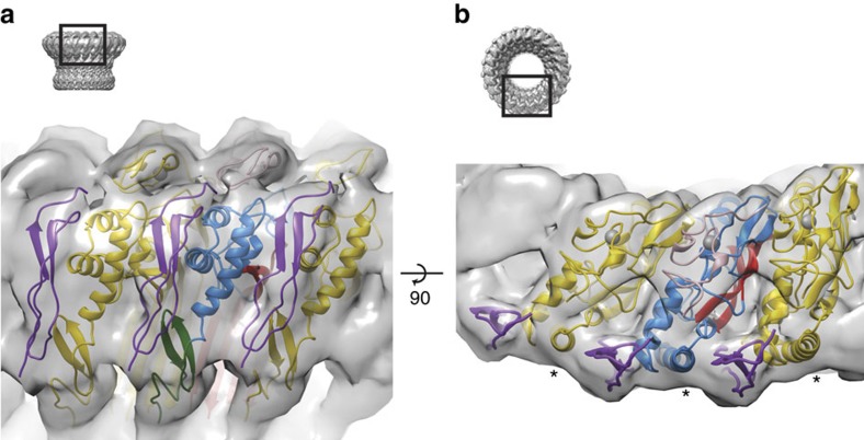
The membrane attack complex (MAC)/perforin-like protein complement component 9 (C9) is the major component of the MAC, a multi-protein complex that forms pores in the membrane of target pathogens. In contrast to homologous proteins such as perforin and the cholesterol-dependent cytolysins (CDCs), all of which require the membrane for oligomerisation, C9 assembles directly onto the nascent MAC from solution. However, the molecular mechanism of MAC assembly remains to be understood. Here we present the 8 Å cryo-EM structure of a soluble form of the poly-C9 component of the MAC. These data reveal a 22-fold symmetrical arrangement of C9 molecules that yield an 88-strand pore-forming β-barrel. The N-terminal thrombospondin-1 (TSP1) domain forms an unexpectedly extensive part of the oligomerisation interface, thus likely facilitating solution-based assembly. These TSP1 interactions may also explain how additional C9 subunits can be recruited to the growing MAC subsequent to membrane insertion.
Figures


References
-
- Kaufmann S. H. E. Immunology's foundation: the 100-year anniversary of the Nobel Prize to Paul Ehrlich and Elie Metchnikoff. Nat. Immunol. 9, 705–712 (2008). - PubMed
-
- Walport M. J. Advances in immunology: complement (first of two parts). N. Engl. J. Med. 344, 1058–1066 (2001). - PubMed
-
- Podack E. R. Molecular mechanisms of cytolysis by complement and by cytolytic lymphocytes. J. Cell Biochem. 30, 133–170 (1986). - PubMed
-
- Tschopp J., Muller-Eberhard H. J. & Podack E. R. Formation of transmembrane tubules by spontaneous polymerization of the hydrophilic complement protein C9. Nature 298, 534–538 (1982). - PubMed
Publication types
MeSH terms
Substances
Grants and funding
LinkOut - more resources
Full Text Sources
Other Literature Sources
Molecular Biology Databases
Research Materials
Miscellaneous
