. 2016 Feb;6(2):150202.
doi: 10.1098/rsob.150202.

Pharmacological removal of serum amyloid P component from intracerebral plaques and cerebrovascular Aβ amyloid deposits in vivo

Affiliations

Pharmacological removal of serum amyloid P component from intracerebral plaques and cerebrovascular Aβ amyloid deposits in vivo

Raya Al-Shawi et al. Open Biol. 2016 Feb.

Abstract

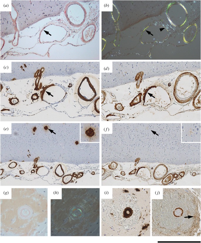
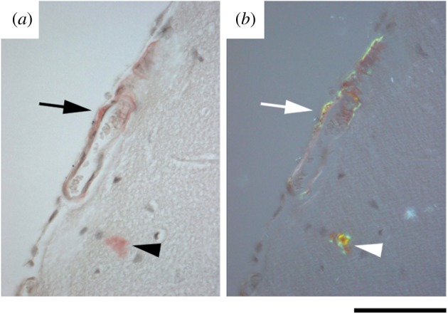
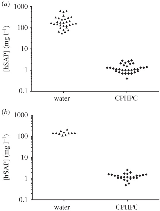
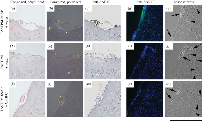
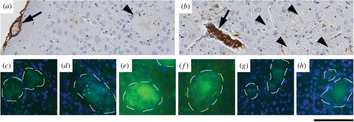
Human amyloid deposits always contain the normal plasma protein serum amyloid P component (SAP), owing to its avid but reversible binding to all amyloid fibrils, including the amyloid β (Aβ) fibrils in the cerebral parenchyma plaques and cerebrovascular amyloid deposits of Alzheimer's disease (AD) and cerebral amyloid angiopathy (CAA). SAP promotes amyloid fibril formation in vitro, contributes to persistence of amyloid in vivo and is also itself directly toxic to cerebral neurons. We therefore developed (R)-1-[6-[(R)-2-carboxy-pyrrolidin-1-yl]-6-oxo-hexanoyl]pyrrolidine-2-carboxylic acid (CPHPC), a drug that removes SAP from the blood, and thereby also from the cerebrospinal fluid (CSF), in patients with AD. Here we report that, after introduction of transgenic human SAP expression in the TASTPM double transgenic mouse model of AD, all the amyloid deposits contained human SAP. Depletion of circulating human SAP by CPHPC administration in these mice removed all detectable human SAP from both the intracerebral and cerebrovascular amyloid. The demonstration that removal of SAP from the blood and CSF also removes it from these amyloid deposits crucially validates the strategy of the forthcoming 'Depletion of serum amyloid P component in Alzheimer's disease (DESPIAD)' clinical trial of CPHPC. The results also strongly support clinical testing of CPHPC in patients with CAA.

Keywords: Alzheimer's disease; Aβ amyloid; CPHPC; cerebral amyloid angiopathy; serum amyloid P component.


Figures

Figure 1.
Figure 1.
Localization of SAP in human CAA. (a,b) Congo red stained section viewed with bright field illumination (a) and with intense crossed polarized light (b), showing abundant amyloid in blood vessel walls. The arrowhead in (b) shows the white birefringence of collagen often seen in normal blood vessels, in contrast to the green birefringence of Congo red stained amyloid. (c,d) Immunoperoxidase staining for Aβ (c) and SAP (d), showing strong immunoreactivity in vessel walls. In one vessel, the amyloid is mostly confined to one segment of the vessel wall (arrow in ad). (e,f) Lower magnification images of the sections shown in (c,d), showing amyloid plaques which stain strongly for Aβ and very weakly for SAP. Higher magnification images of the arrowed plaque are inset. (gj) Sections of an amyloid-containing blood vessel surrounded by blood. (g,h) Congo red stained section viewed with bright field illumination (g) and with intense cross polarized light (h). (i,j) Immunostaining for Aβ and SAP, respectively. The arrow in (j) shows diffuse staining in adjacent neural tissue, not associated with amyloid, likely due to a microbleed. Scale bars, (ad), 250 µm; (e,f), 500 µm; (gj), 250 µm.
Figure 2.
Figure 2.
Cerebrovascular amyloid in TASTPM mice. Unfixed frozen brain sections from six-month-old female TASTPM mouse stained with Congo red showing cerebrovascular amyloid (arrow) and cerebral plaque (arrowhead). (a) Bright field illumination; (b) intense cross polarized light showing pathognomonic green birefringence of Congo red bound to amyloid. Scale bar, 150 µm.
Figure 3.
Figure 3.
Human SAP expression in transgenic mice, and depletion by CPHPC. Human SAP concentrations in sera of individual transgenic mice, while on water alone or water containing 5 mg ml−1 CPHPC. (a) Line 38 human SAP transgenic C57BL/6 mice; (b) line 38 human SAP transgenic TASTPM mice. Results are from males and females with no apparent differences between sexes.
Figure 4.
Figure 4.
Depletion of human SAP from cerebrovascular amyloid by CPHPC treatment of transgenic mice. Sections of formalin fixed wax embedded cerebral cortex from 20-month-old mice stained with Congo red and viewed in bright field (a,f,k) or intense cross polarized illumination (b,g,l), and also immunoperoxidase (IP) stained with anti-human SAP antibodies (c,h,m). Unfixed frozen sections from the same tissues stained by immunofluorescence (IF) with anti-human SAP antibodies (green) and bisbenzimide counterstain (blue) (d,i,n) and also viewed in phase contrast (e,j,o). (ae) Human SAP transgenic TASTPM mouse, (fj) TASTPM control mouse, (ko) CPHPC-treated human SAP transgenic TASTPM mouse. Amyloid-containing blood vessels are labelled with arrows, and example amyloid plaques are indicated by arrowheads. Tissues in (ac), (fh), (km) are from males and (d,e,i,j,n,o) from female mice.
Figure 5.
Figure 5.
Depletion of human SAP from cerebral plaques by CPHPC treatment of transgenic mice. (a,b) Sections of formalin fixed wax embedded cerebral cortex from 20-month-old human SAP transgenic male TASTPM mice, immunoperoxidase stained with anti-human SAP antibodies, showing intense staining of vascular amyloid (arrows) and very weak staining of intracerebral plaques (arrowheads). (ch) Amyloid plaques (outlined) in unfixed frozen sections of cerebral cortex from 20-month-old female mice stained with fluorescent labelled anti-human SAP antibodies (green). (c,d) TASTPM control; (e,f) human SAP transgenic TASTPM; (g,h) CPHPC-treated human SAP transgenic TASTPM. The green channel exposures used for panels (ch) were 10 times longer than for figure 4d,i,n. At the exposure that demonstrated SAP in the cerebrovascular amyloid in figure 4, there was no signal from the intracerebral plaques. Scale bars, (a,b), 100 µm; (ch), 150 µm.

Similar articles

Cited by

References

    1. Glenner GG, Wong CW. 1984. Alzheimer's disease and Down's syndrome: sharing of a unique cerebrovascular amyloid fibril protein. Biochem. Biophys. Res. Commun. 122, 1131–1135. (doi:10.1016/0006-291X(84)91209-9) - DOI - PubMed
    1. De Strooper B, König G. 1999. Alzheimer's disease. A firm base for drug development. Nature 402, 471–472. (doi:10.1038/44973) - DOI - PubMed
    1. Brunkan AL, Goate AM. 2005. Presenilin function and γ-secretase activity. J. Neurochem. 93, 769–792. (doi:10.1111/j.1471-4159.2005.03099.x) - DOI - PubMed
    1. Bohm C, Chen F, Sevalle J, Qamar S, Dodd R, Li Y, Schmitt-Ulms G, Fraser PE, St George-Hyslop PH. 2015. Current and future implications of basic and translational research on amyloid-β peptide production and removal pathways. Mol. Cell. Neurosci. 66, 3–11. (doi:10.1016/j.mcn.2015.02.016) - DOI - PMC - PubMed
    1. Schneider LS, et al. 2014. Clinical trials and late-stage drug development for Alzheimer's disease: an appraisal from 1984 to 2014. J. Intern. Med. 275, 251–283. (doi:10.1111/joim.12191) - DOI - PMC - PubMed

Publication types

MeSH terms

Substances