

Case Reports
doi: 10.3904/kjim.2015.107.
Epub 2016 Feb 3.
Acute cor pulmonale due to pulmonary tumor thrombotic microangiopathy in two patients with breast cancer
Affiliations
- PMID: 26842105
- PMCID: PMC5214717
- DOI: 10.3904/kjim.2015.107
Item in Clipboard
Case Reports
Acute cor pulmonale due to pulmonary tumor thrombotic microangiopathy in two patients with breast cancer
Korean J Intern Med.
2017 Jan.
No abstract available
Keywords: Breast neoplasms; Pulmonary heart disease; Thrombi.
Conflict of interest statement
No potential conflict of interest relevant to this article was reported.
Figures
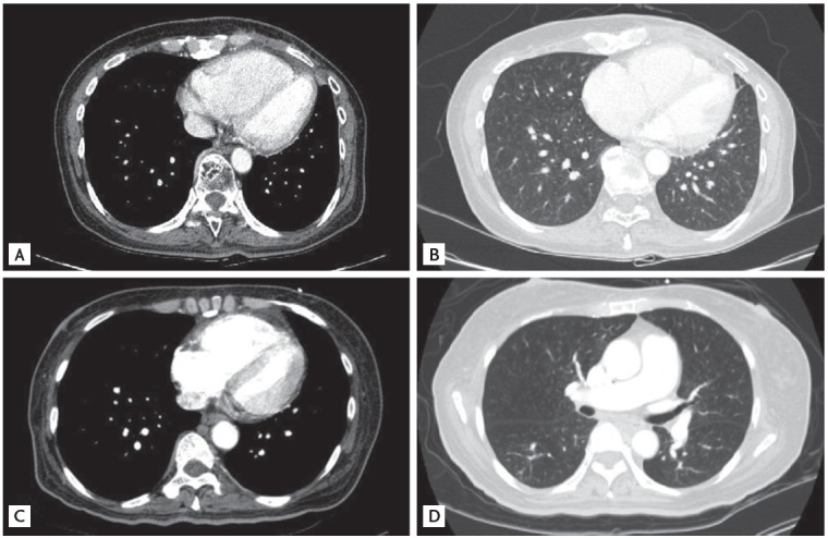
Computed tomography showed multiple centrilobular nodules with tree-in-bud appearances in cases 1, 2. A mediastinal setting showed a dilated right ventricle and a compressed D-shaped left ventricle in cases 1 and 2, respectively (A, C). A lung setting showed multiple ill-defined centrilobular nodules with tree-in-bud appearances in cases 1 and 2, respectively (B, D).
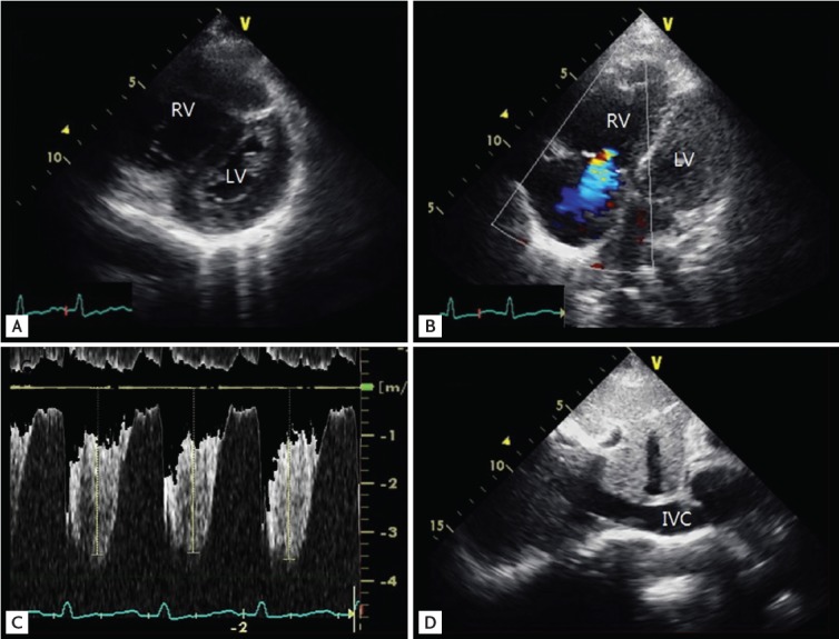
Echocardiography in case 2. (A) Parasternal short axis view, mid-ventricular plane on transthoracic echocardiography reveals a D-shaped LV throughout the systolic and diastolic period. (B) The 2-dimensional and color Doppler comparative focused image of the apical 4-chamber view showed mild-to-moderate tricuspid regurgitation. (C) The peak tricuspid regurgitation velocity was 3.7 m/sec, pressure gradient 55 mmHg, indicating pulmonary hypertension. (D) The dilated inferior vena cava (IVC) diameter in case 1 was 21 mm without inspiratory collapse. RV, right ventricle; LV, left ventricle.
References
-
- von Herbay A, Illes A, Waldherr R, Otto HF. Pulmonary tumor thrombotic microangiopathy with pulmonary hypertension. Cancer. 1990;66:587–592. - PubMed
-
- Montero A, Vidaller A, Mitjavila F, Chivite D, Pujol R. Microscopic pulmonary tumoral embolism and subacute cor pulmonale as the first clinical signs of cancer. Acta Oncol. 1999;38:1116–1118. - PubMed
-
- Uruga H, Fujii T, Kurosaki A, et al. Pulmonary tumor thrombotic microangiopathy: a clinical analysis of 30 autopsy cases. Intern Med. 2013;52:1317–1323. - PubMed
-
- Miyano S, Izumi S, Takeda Y, et al. Pulmonary tumor thrombotic microangiopathy. J Clin Oncol. 2007;25:597–599. - PubMed
Publication types
MeSH terms
LinkOut - more resources
Full Text Sources
Other Literature Sources
Medical