Epithelioid angiosarcoma arising in schwannoma of the kidney: report of the first case and review of the literature
- PMID: 26842370
- PMCID: PMC4739400
- DOI: 10.1186/s12957-016-0789-5
Epithelioid angiosarcoma arising in schwannoma of the kidney: report of the first case and review of the literature
Abstract
Background: Schwannoma and angiosarcoma are infrequent pathologies that have been rarely reported in the kidney. Angiosarcoma is an uncommon malignant tumor presenting a recognizable vascular differentiation. It can develop in any site but the most common locations include the skin, soft tissues, breast, bone, liver, and spleen while renal localization has been very rarely reported in the literature. Schwannoma is a benign peripheral nerve sheath tumor composed of cells with the immunophenotype and ultrastructural features of differentiated Schwann cells. It has a wide anatomical distribution but the most frequent locations include subcutaneous tissues of the extremities and the head and neck region and the retroperitoneal and mediastinal soft tissues. The occurrence of an angiosarcoma in a pre-existing schwannoma is an extremely rare event with <20 cases reported in worldwide literature. In the present study, a renal case of angiosarcoma arising in schwannoma is presented with a detailed review of the pertinent literature.
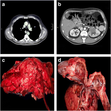
Case presentation: A 56-year-old man was admitted with a few days history of lower back pain and hematuria. Abdominal ultrasound showed a mass inside the left renal medulla. Subsequent imaging investigations with computed tomography and magnetic resonance confirmed the presence of the lesion and showed a pulmonary metastasis.
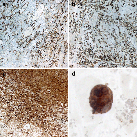
Conclusions: The final histopathological examination led to the diagnosis of epithelioid angiosarcoma arising in a schwannoma. The patient came to death a few months later due to a massive hemothorax. To the best of our knowledge, the present is the first case of an angiosarcoma arising in a schwannoma of the kidney.
Figures


References
-
- Fletcher CDM, Bridge JA, Hogendoorn PCW, Mertens F. WHO classification of tumours of soft tissue and bone, 4th edition. International Agency for Research on Cancer (IARC); Philadephia, U.S.A., 2013.
-
- Eldridge R. Central neurofibromatosis with bilateral acoustic neuroma. Adv Neurol. 1981;29:57–65. - PubMed
Publication types
MeSH terms
LinkOut - more resources
Full Text Sources
Other Literature Sources
Medical

