

Comparison of the Automated cobas u 701 Urine Microscopy and UF-1000i Flow Cytometry Systems and Manual Microscopy in the Examination of Urine Sediments
- PMID: 26842372
- PMCID: PMC6807231
- DOI: 10.1002/jcla.21919
Comparison of the Automated cobas u 701 Urine Microscopy and UF-1000i Flow Cytometry Systems and Manual Microscopy in the Examination of Urine Sediments
Abstract
Background: The cobas u 701, a new automated image-based urine sediment analyzer, was introduced recently. In this study, we compared its performance with that of UF-1000i flow cytometry and manual microscopy in the examination of urine sediments.
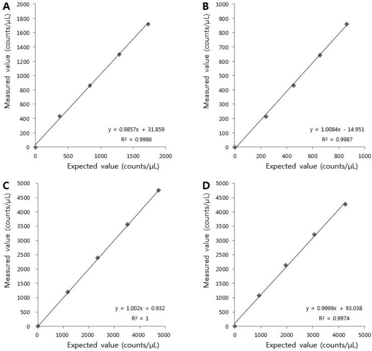
Methods: Precision, linearity, and carry-over were determined for the two urine sediment analyzers. For a comparison of the method, 300 urine samples were examined by the automated analyzers and by manual microscopy using a KOVA chamber.
Results: Within-run coefficients of variation (CVs) for the control materials were 7.0-8.8% and 1.7-5.7% for the cobas u 701 and UF-1000i systems, respectively. Between-run CVs were 8.5-9.8% and 2.7-5.4%, respectively. Both instruments showed good linearity and negligible carry-over. For red blood cells (RBC), white blood cells (WBC), and epithelial cells (EPI), the overall concordance rates within one grade of difference among the three methods were good (78.6-86.0%, 88.7-93.8%, and 81.3-90.7%, respectively). The concordance rate for casts was poor (66.5-68.9%).
Conclusion: Compared with manual microscopy, the two automated sediment analyzers tested in this study showed satisfactory analytical performances for RBC, WBC, and EPI. However, for other urine sediment particles confirmation by visual microscopy is still required.
Keywords: UF-1000i; automated urine sediment analyzer; cobas u 701; urine microscopy; urine sediment.
© 2016 Wiley Periodicals, Inc.
Conflict of interest statement
No potential conflict of interest relevant to this article were reported.
Figures
References
-
- McPherson RA, Ben‐Ezra J. Basic examination of urine McPherson RA, Pincus MR. (eds.). Henry's Clinical Diagnosis and Management by Laboratory Methods, 22nd edition, Philadelphia: Saunders; 2011. p 445–479.
-
- Zaman Z, Fogazzi GB, Garigali G, Croci MD, Bayer G, Kranicz T. Urine sediment analysis: Analytical and diagnostic performance of sediMAX®—A new automated microscopy image‐based urine sediment analyser. Clin Chim Acta 2010;411:147–154. - PubMed
-
- Manoni F, Tinello A, Fornasiero L, et al. Urine particle evaluation: A comparison between the UF‐1000i and quantitative microscopy. Clin Chem Lab Med 2010;48:1107–1111. - PubMed
Publication types
MeSH terms
LinkOut - more resources
Full Text Sources
Other Literature Sources
Medical
