. 2016 Feb;9(1):51-9.
doi: 10.1111/cts.12366. Epub 2016 Feb 4.

ABCC4 Is a Determinant of Cytarabine-Induced Cytotoxicity and Myelosuppression

Affiliations

ABCC4 Is a Determinant of Cytarabine-Induced Cytotoxicity and Myelosuppression

C D Drenberg et al. Clin Transl Sci. 2016 Feb.

Abstract

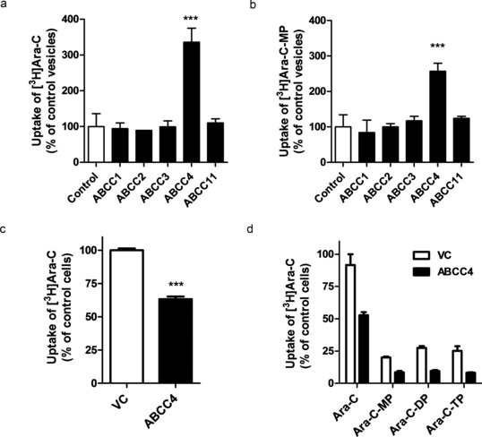
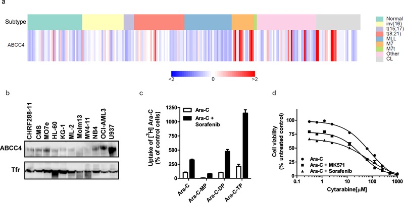
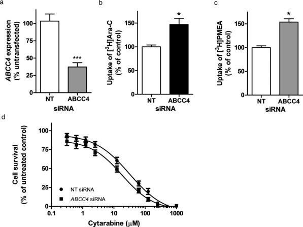
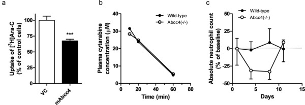
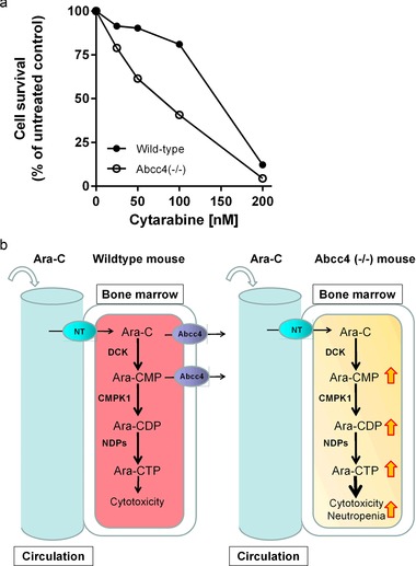
Resistance to cytarabine remains a major challenge in the treatment of acute myeloid leukemia (AML). Based on previous studies implicating ABCC4/MRP4 in the transport of nucleosides, we hypothesized that cytarabine is sensitive to ABCC4-mediated efflux, thereby decreasing its cytotoxic response against AML blasts. The uptake of cytarabine and its monophosphate metabolite was found to be facilitated in ABCC4-expressing vesicles and intracellular retention was significantly impaired by overexpression of human ABCC4 or mouse Abcc4 (P < 0.05). ABCC4 was expressed highly in AML primary blasts and cell lines, and cytotoxicity of cytarabine in cells was increased in the presence of the ABCC4 inhibitors MK571 or sorafenib, as well as after ABCC4 siRNA. In Abcc4-null mice, cytarabine-induced hematological toxicity was enhanced and ex vivo colony-forming assays showed that Abcc4-deficiency sensitized myeloid progenitors to cytarabine. Collectively, these studies demonstrate that ABCC4 plays a protective role against cytarabine-mediated insults in leukemic and host myeloid cells.


Figures

Figure 1
Figure 1
Transport of cytarabine (Ara‐C) and cytarabine‐monophosphate (Ara‐CMP) by ABCC4. Uptake of (a) Ara‐C and (b) Ara‐CMP in vesicles expressing the indicated ABCC transporters. Vesicles were incubated with 1.25 μM Ara‐C or 50 μM Ara‐CMP for 5 minutes. Results were normalized to ATP‐independent transport and shown as percentage of uptake in control vesicles. Intracellular accumulation of (c) Ara‐C and (d) Ara‐C and phosphorylated metabolites in Saos‐2 cells transfected with human ABCC4 (ABCC4). Cells were incubated with 1.25 μM Ara‐C for 2 hours (in C) or 4 hours (in D). Ara‐C, Ara‐C‐MP, Ara‐C‐diphosphate (Ara‐C‐DP), and Ara‐C triphosphate (Ara‐C‐TP) were measured in cell lysates by HPLC and radioactivity was determined by liquid scintillation counting. Results are shown as percentage of uptake in control cells transfected with empty vector (VC). Controls were set at a value of 100% and data are presented as mean (bars) and SD (error bars) of two to three independent experiments (four to six replicates). ***P < 0.001 vs. control.
Figure 2
Figure 2
ABCC4 is expressed in childhood primary AML blast samples and human AML cell lines and contributes to cytarabine (Ara‐C)‐induced cytotoxicity. (a) ABCC4 gene expression in primary samples and cell lines assessed by microarray analysis. Each column represents normal bone marrow (normal), an individual primary sample, or cell line (CL); columns are categorized by cytogenetic AML subtypes of prognostic relevance (data from Ref. 12). (b) Protein expression of ABCC4 in AML cell lines. Cellular membrane protein (20 μg) was loaded in each lane and protein expression was assessed by western blotting using an antibody to ABCC4 and transferrin receptor (Tfr) as a loading control. (c) Accumulation of Ara‐C and phosphorylated metabolites in OCI‐AML3 cells in the absence or presence of sorafenib. Cells were treated with 1.25 μM Ara‐C alone or in combination with 5 μM sorafenib for 2 hours. Ara‐C, Ara‐C monophosphate (Ara‐C‐MP), Ara‐C‐diphosphate (Ara‐C‐DP), and Ara‐C triphosphate (Ara‐C‐TP) were measured in cell lysates by HPLC and radioactivity was determined by liquid scintillation counting. Accumulation of Ara‐C in the absence of sorafenib was set at a value of 100% (control) and data are shown as a percentage of uptake in controls and presented as mean (bars) and SD (error bars) of two independent experiments (four replicates). (d) Cytarabine‐induced cytotoxicity in the absence or presence of MK571 or sorafenib in OCI‐AML3 cells. Cells were incubated with increasing concentrations of Ara‐C alone and with 100 μM MK571 or 10 μM sorafenib. Data are presented as mean (bars) and SD (error bars) of three independent experiments (24 replicates).
Figure 3
Figure 3
Knockdown of ABCC4 in OCI‐AML3 cells increases cellular accumulation of cytarabine, and enhances cytotoxicity to cytarabine. (a) ABCC4 mRNA expression in OCI‐AML3 cells transfected with 3 μg of ABCC4 siRNA or a nontargeting (NT) control siRNA. ABCC4 expression was determined by real‐time PCR. Results are shown as a percentage of untransfected normal cells, with values in cells transfected with control NT siRNA set at 100%. Data are representative of one of two independent experiments performed in triplicate. Cellular accumulation of (b) cytarabine (Ara‐C) and (c) PMEA were increased after ABCC4 knockdown by siRNA. The results are accumulation at 2 hours, with cells transfected with control NT siRNA set at 100%. PMEA was used as a positive control substrate. Results represent an average of two to three independent experiments with two to six replicates. (d) Cytarabine cytotoxicity in OCI‐AML3 cells after ABCC4 siRNA knockdown. Data are presented as mean of two to three independent experiments (eight replicates). *P < 0.05 vs. control; ***P < 0.001 vs. control.
Figure 4
Figure 4
Cytarabine‐induced myelosuppression is enhanced in Abcc4(–/–) mice. (a) Intracellular accumulation of cytarabine (Ara‐C) in Saos‐2 cells transfected with mouse Abcc4 (mAbcc4). Cells were incubated with 5 μM Ara‐C for 2 hours and the results are shown as the percentage of control cells transfected with empty vector (VC) set at a value of 100%. Data are presented as mean (bars) and SD (error bars) of three independent experiments (nine replicates). ***P < 0.001 vs. control. (b) Plasma concentrations of cytarabine are unaffected by Abcc4‐deficiency in mice after i.p. injection of 12.5 mg/kg of cytarabine. (c) Mice were treated with 12.5 mg/kg of cytarabine once daily for 5 days, and neutrophil counts were monitored and shown as percentage change from baseline.
Figure 5
Figure 5
Abcc4‐deficiency sensitizes myeloid progenitor cells to cytarabine. (a) c‐Kit+, Sca‐1+, and Lin (KSL) cells from bone marrow of wildtype and Abcc4(–/–) mice were cultured in methyl cellulose culture medium with increasing concentrations of cytarabine. Granulocyte macrophage colony‐forming cell assay was determined after 7 days of culture. Duplicate plates were cultured at each drug concentration for two independent experiments. Data represent the percentage of viable colonies at different concentrations. (b) Theoretical influence of Abcc4 knockout on cytarabine‐induced cytotoxicity and myelosuppression. A nucleotide transporter facilitates cytarabine entry into the cell, where it is rapidly metabolized into its active triphosphate form through a series of enzymatic reactions. In wildtype cells, Abcc4 can exert a protective function by facilitating export of parent compound and the Ara‐CMP metabolite. Knockout of Abcc4 in mice promotes intracellular accumulation of cytarabine and its metabolites, resulting in enhanced cytotoxicity and myelosuppression. Alternatively, Abcc4 function may be inhibited chemically (sorafenib, MK571) or through genetic variation. The black arrows indicate the direction of transport. The red/yellow arrows indicate the accumulation of cytarabine and metabolites that occurs when Abcc4 is inhibited. Ara‐C, cytarabine; Ara‐CMP, cytarabine monophosphate; Ara‐CDP, cytarabine diphosphate; Ara‐CTP, cytarabine triphosphate; CMPK1, cytidine monophosphate kinase 1; DCK, deoxycytidine kinase; NDPs, nucleotide diphosphate kinases; NT, nucleotide transporter.

References

    1. Borst, P. , de Wolf, C. & van de Wetering, K. Multidrug resistance‐associated proteins 3, 4, and 5. Pflugers Arch. 453, 661–673 (2007). - PubMed
    1. Peng, X.X. et al Up‐regulation of MRP4 and down‐regulation of influx transporters in human leukemic cells with acquired resistance to 6‐mercaptopurine. Leuk. Res. 32, 799–809 (2008). - PubMed
    1. Liu, B. , Zhao, L. , Ma, H. , Zhang, W. & Jin, Y. Knockdown of MRP4 by lentivirus‐mediated siRNA improves sensitivity to adriamycin in adriamycin‐resistant acute myeloid leukemia cells. Chin. Sci. Bull. 57, 90–97 (2012).
    1. Copsel, S. et al Multidrug resistance protein 4 (MRP4/ABCC4) regulates cAMP cellular levels and controls human leukemia cell proliferation and differentiation. J. Biol. Chem. 286, 6979–6988 (2011). - PMC - PubMed
    1. Oevermann, L. et al Hematopoietic stem cell differentiation affects expression and function of MRP4 (ABCC4), a transport protein for signaling molecules and drugs. Int. J. Cancer 124, 2303–2311 (2009). - PubMed

Publication types

MeSH terms

LinkOut - more resources