

Small-molecule CFTR activators increase tear secretion and prevent experimental dry eye disease
- PMID: 26842854
- PMCID: PMC4836376
- DOI: 10.1096/fj.201500180
Small-molecule CFTR activators increase tear secretion and prevent experimental dry eye disease
Abstract
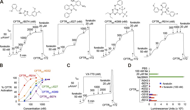
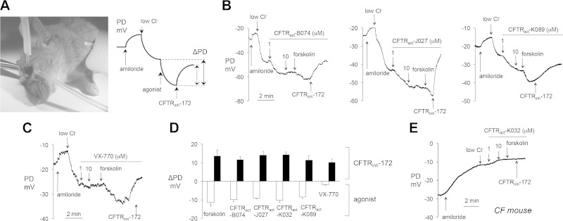
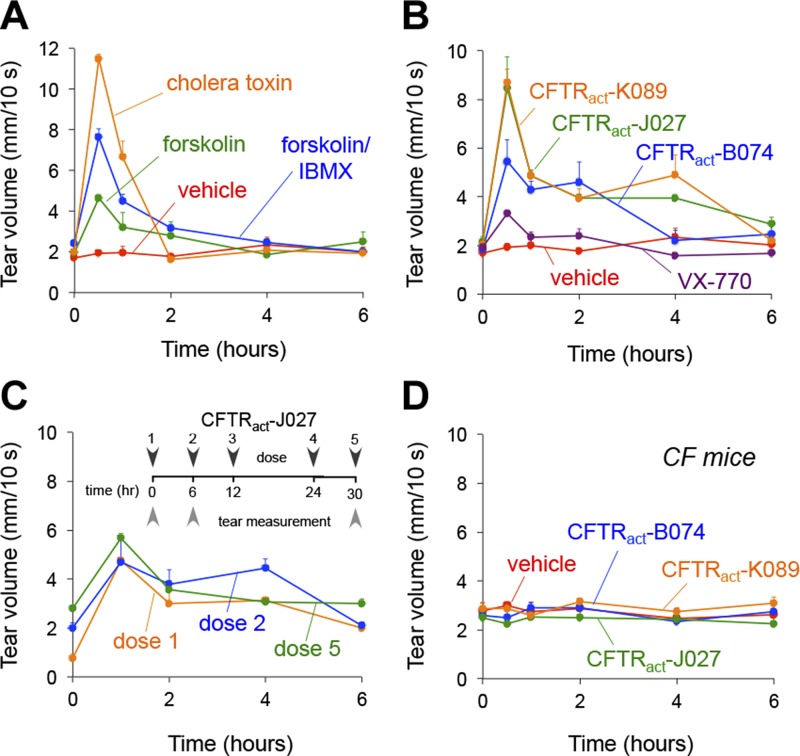
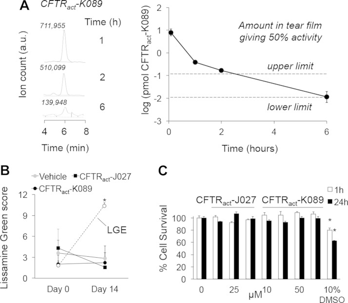
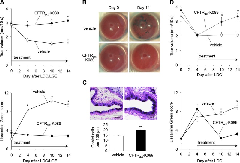
Dry eye disorders, including Sjögren's syndrome, constitute a common problem in the aging population, with limited effective therapeutic options available. The cAMP-activated Cl(-) channel cystic fibrosis transmembrane conductance regulator (CFTR) is a major prosecretory channel at the ocular surface. We investigated whether compounds that target CFTR can correct the abnormal tear film in dry eye. Small-molecule activators of human wild-type CFTR identified by high-throughput screening were evaluated in cell culture and in vivo assays, to select compounds that stimulate Cl(-)-driven fluid secretion across the ocular surface in mice. An aminophenyl-1,3,5-triazine, CFTRact-K089, fully activated CFTR in cell cultures with EC50 ∼250 nM and produced an ∼8.5 mV hyperpolarization in ocular surface potential difference. When delivered topically, CFTRact-K089 doubled basal tear volume for 4 h and had no effect in CF mice. CFTRact-K089 showed sustained tear film bioavailability without detectable systemic absorption. In a mouse model of aqueous-deficient dry eye produced by lacrimal ablation, topical administration of 0.1 nmol CFTRact-K089 3 times daily restored tear volume to basal levels, preventing corneal epithelial disruption when initiated at the time of surgery and reversing it when started after development of dry eye. Our results support the potential utility of CFTR-targeted activators as a novel prosecretory treatment for dry eye.-Flores, A. M., Casey, S. D., Felix, C. M., Phuan, P. W., Verkman, A. S., Levin, M. H. Small-molecule CFTR activators increase tear secretion and prevent experimental dry eye disease.
Keywords: chloride channels; conjunctiva; cornea; ocular surface.
© FASEB.
Figures
Similar articles
-
Nanomolar Potency Aminophenyltriazine CFTR Activator Reverses Corneal Epithelial Injury in a Mouse Model of Dry Eye.J Ocul Pharmacol Ther. 2020 Apr;36(3):147-153. doi: 10.1089/jop.2019.0087. Epub 2020 Jan 14. J Ocul Pharmacol Ther. 2020. PMID: 31934802 Free PMC article.
-
Nanomolar-Potency Aminophenyl-1,3,5-triazine Activators of the Cystic Fibrosis Transmembrane Conductance Regulator (CFTR) Chloride Channel for Prosecretory Therapy of Dry Eye Diseases.J Med Chem. 2017 Feb 9;60(3):1210-1218. doi: 10.1021/acs.jmedchem.6b01792. Epub 2017 Jan 31. J Med Chem. 2017. PMID: 28099811 Free PMC article.
-
Pro-Secretory Activity and Pharmacology in Rabbits of an Aminophenyl-1,3,5-Triazine CFTR Activator for Dry Eye Disorders.Invest Ophthalmol Vis Sci. 2017 Sep 1;58(11):4506-4513. doi: 10.1167/iovs.17-22525. Invest Ophthalmol Vis Sci. 2017. PMID: 28873176 Free PMC article.
-
Overview of CFTR activators and their recent studies for dry eye disease: a review.RSC Med Chem. 2023 Sep 25;14(12):2459-2472. doi: 10.1039/d3md00448a. eCollection 2023 Dec 13. RSC Med Chem. 2023. PMID: 38107177 Free PMC article. Review.
-
Differential diagnosis of dry eye conditions.Adv Dent Res. 1996 Apr;10(1):9-12. doi: 10.1177/08959374960100011801. Adv Dent Res. 1996. PMID: 8934916 Review.
Cited by
-
Benzopyrimido-pyrrolo-oxazine-dione CFTR inhibitor (R)-BPO-27 for antisecretory therapy of diarrheas caused by bacterial enterotoxins.FASEB J. 2017 Feb;31(2):751-760. doi: 10.1096/fj.201600891R. Epub 2016 Nov 8. FASEB J. 2017. PMID: 27871064 Free PMC article.
-
Ion channels in dry eye disease.Indian J Ophthalmol. 2023 Apr;71(4):1215-1226. doi: 10.4103/IJO.IJO_3020_22. Indian J Ophthalmol. 2023. PMID: 37026252 Free PMC article. Review.
-
Nanomolar Potency Aminophenyltriazine CFTR Activator Reverses Corneal Epithelial Injury in a Mouse Model of Dry Eye.J Ocul Pharmacol Ther. 2020 Apr;36(3):147-153. doi: 10.1089/jop.2019.0087. Epub 2020 Jan 14. J Ocul Pharmacol Ther. 2020. PMID: 31934802 Free PMC article.
-
Novel Insight Into the Role of CFTR in Lacrimal Gland Duct Function in Mice.Invest Ophthalmol Vis Sci. 2018 Jan 1;59(1):54-62. doi: 10.1167/iovs.17-22533. Invest Ophthalmol Vis Sci. 2018. PMID: 29305607 Free PMC article.
-
Cellular mechanism for herbal medicine Junchoto to facilitate intestinal Cl-/water secretion that involves cAMP-dependent activation of CFTR.J Nat Med. 2018 Jun;72(3):694-705. doi: 10.1007/s11418-018-1207-9. Epub 2018 Mar 22. J Nat Med. 2018. PMID: 29569221 Free PMC article.
References
-
- Dry Eye WorkShop (DEWS) Definition and Classification Subcommittee (2007) The definition and classification of dry eye disease: report of the Definition and Classification Subcommittee of the International Dry Eye WorkShop 2007 Ocul. Surf. 5, 75–92 - PubMed
-
- Schaumberg D. A., Sullivan D. A., Buring J. E., Dana M. R. (2003) Prevalence of dry eye syndrome among US women. Am. J. Ophthalmol. 136, 318–326 - PubMed
-
- Yu J., Asche C. V., Fairchild C. J. (2011) The economic burden of dry eye disease in the United States: a decision tree analysis. Cornea 30, 379–387 - PubMed
-
- Alves M., Fonseca E. C., Alves M. F., Malki L. T., Arruda G. V., Reinach P. S., Rocha E. M. (2013) Dry eye disease treatment: a systematic review of published trials and a critical appraisal of therapeutic strategies. Ocul. Surf. 11, 181–192 - PubMed
MeSH terms
Substances
Grants and funding
LinkOut - more resources
Full Text Sources
Other Literature Sources
Medical
