. 2016 May;17(5):475-86.
doi: 10.1111/tra.12385. Epub 2016 Mar 28.

Engineered Tug-of-War Between Kinesin and Dynein Controls Direction of Microtubule Based Transport In Vivo

Affiliations

Engineered Tug-of-War Between Kinesin and Dynein Controls Direction of Microtubule Based Transport In Vivo

Karim Rezaul et al. Traffic. 2016 May.

Abstract

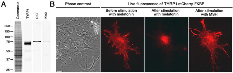
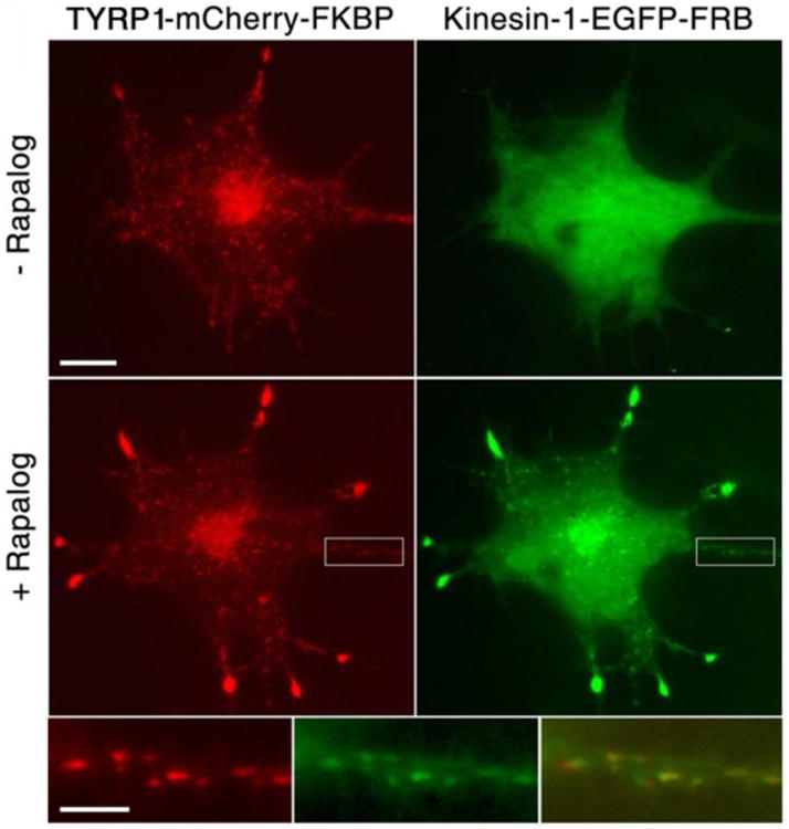
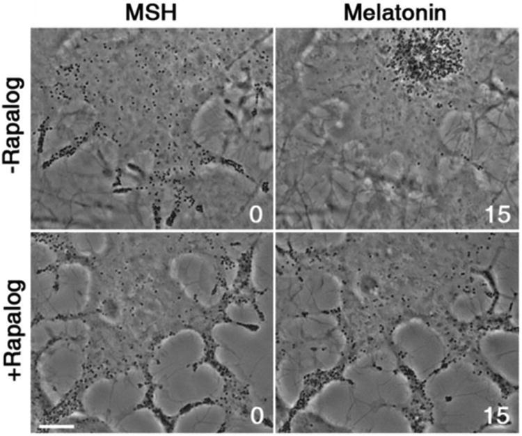
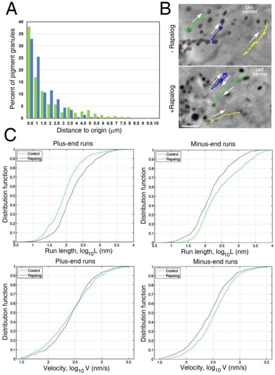
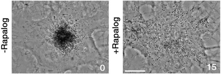
Bidirectional transport of membrane organelles along microtubules (MTs) is driven by plus-end directed kinesins and minus-end directed dynein bound to the same cargo. Activities of opposing MT motors produce bidirectional movement of membrane organelles and cytoplasmic particles along MT transport tracks. Directionality of MT-based transport might be controlled by a protein complex that determines which motor type is active at any given moment of time, or determined by the outcome of a tug-of-war between MT motors dragging cargo organelles in opposite directions. However, evidence in support of each mechanisms of regulation is based mostly on the results of theoretical analyses or indirect experimental data. Here, we test whether the direction of movement of membrane organelles in vivo can be controlled by the tug-of-war between opposing MT motors alone, by attaching a large number of kinesin-1 motors to organelles transported by dynein to minus-ends of MTs. We find that recruitment of kinesin significantly reduces the length and velocity of minus-end-directed dynein-dependent MT runs, leading to a reversal of the overall direction of dynein-driven organelles in vivo. Therefore, in the absence of external regulators tug-of-war between opposing MT motors alone is sufficient to determine the directionality of MT transport in vivo.

Keywords: dynein; intracellular transport; kinesin; melanophore; pigment granule.


Figures

Figure 1
Figure 1
TRP-1 binds pigment granules in melanophores. (A) Immunoblots of preparations of pigment granules probed with antibodies against TRP-1 or subunits of motor proteins involved in pigment granule transport; coomassie-stained gel of a preparation of pigment granules (left), immunoblots of pigment granule preparations with antibodies raised against TRP-1 (middle left), dynein intermediate chain (DIC; middle right), and A subunit of kinesin-2 (kin2; right). (B) Phase contrast (left) and fluorescence images of a pigment-free melanophore expressing TRP-1-FKBP-mCherry before (middle left) or after (middle right) stimulation with melatonin, or subsequent treatment with MSH (right); TRP-mCherry-FKBP is localized to fluorescent dots that accumulate in the cell center or disperse throughout the cytoplasm after treatment of cells with melatonin and MSH, respectively, as would be expected for pigment granules. Scale bar, 5 μm.
Figure 2
Figure 2
Rapalog treatment recruits kinesin-1-EGFP- FRB to the pigment granules that bound TRP-1-FKBP-mCherry. Top and middle, fluorescence images of melanophores co-expressing TRP-1-FKBP-mCherry (left) and kinesin-1-EGFP- FRB (right) before (top) or after (middle) stimulation with rapalog; bottom, high magnification images (left and middle) and overlay (right) of boxed areas shown in low magnification images. Treatment of melanophores with rapalog leads to acquisition of EGFP fluorescence by the mCherry positive dots, indicating that kinesin-1-EGFP- FRB binds pigment granules. Scale bars, 5 μm (top) and 2 μm (bottom).
Figure 3
Figure 3
Recruitment of kinesin-1 inhibits accumulation of pigment granules in the cell center induced by pigment aggregation signals. Phase contrast images of melanophores co-expressing kinesin-1-EGFP- FRB and TRP-1-FKBP-mCherry treated with MSH (left images) or melatonin (right images) in the absence (top row) or presence (bottom row) of rapalog; rapalog treatment that recruits kinesin-1 to pigment granules inhibits their aggregation in the cell center. Numbers indicate time in minutes. Scale bar, 5 μm.
Figure 4
Figure 4
Recruitment of kinesin-1-EGFP- FRB does not inhibit motility of pigment granules but enhances their plus-end directed movement by increasing the length of plus-end runs and reducing the length and velocity of minus-end runs. (A) Frequency histogram of distances traveled by pigment granules in control (green) or rapalog-treated (blue) cells within 15 s time interval used for recoding of the granule motility; in control cells, pigment granules more frequently travel for longer distances compared to rapalog-treated cells, but rapalog treatment does not increase the fraction of essentially immotile granules that traveled for distances not exceeding 0.5 μm (average diameter of pigment granules), - an indication that recruitment of kinesin-1 does not block pigment granule motility. (B)Motility tracks of pigment granules in control (top) or rapalog-treated (bottom) melanophores co-expressing kinesin-1-EGFP- FRB and TRP-1-FKBP-mCherry and stimulated with melatonin. Rapalog treatment increases the frequency and length of granule runs directed away from the cell center (upper right corner of each image). Color-coded lines indicate tracks of individual granules. Arrows indicate direction of granule movement. Numbers indicate time in minutes. Scale bar, 2 μm. (C) Cumulative distribution function plots that show the probability distribution for lengths (top) or velocities (bottom) of plus-end (left) or minus-end (right) granule runs in control (green) or rapalog-treated (blue) cells coexpressing kinesin-1-EGFP- FRB and TRP-1-FKBP-mCherry; recruitment of kinesin-1-EGFP- FRB increases the probability of longer plus-end granule runs (upper left) without changing the probability distribution of their velocities (lower left), and at the same time increases the probabilities of shorter and slower minus-end runs (upper right and lower right, respectively), - an indication that kinesin-1 interferes with dynein motility.
Figure 5
Figure 5
Recruitment of kinesin-1 reverses direction of pigment granule transport induced by pigment aggregation signals. Phase contrast images of a cell co-expressing kinesin-1-EGFP- FRB and TRP-1-FKBP-mCherry treated with melatonin to aggregate pigment granules before (left) or after (right) treatment with rapalog; rapalog-induced recruitment of kinesin-1 leads to dispersion of pigment granules, and therefore reverses direction of their dynein-dependent movement. Numbers indicate time in minutes. Scale bar, 5 μm.

References

    1. Vale RD. The molecular motor toolbox for intracellular transport. Cell. 2003;112(4):467–480. - PubMed
    1. Hancock WO. Bidirectional cargo transport: moving beyond tug of war. Nature reviews Molecular cell biology. 2014;15(9):615–628. - PMC - PubMed
    1. Jolly AL, Gelfand VI. Bidirectional intracellular transport: utility and mechanism. Biochemical Society transactions. 2011;39(5):1126–1130. - PMC - PubMed
    1. Gross SP. Hither and yon: a review of bi-directional microtubule-based transport. Phys Biol. 2004;1(1-2):R1–11. - PubMed
    1. Welte MA. Bidirectional transport along microtubules. Curr Biol. 2004;14(13):R525–537. - PubMed

Publication types

LinkOut - more resources