. 2016 Apr;89(4):457-66.
doi: 10.1124/mol.115.101808. Epub 2016 Feb 3.

Evidence for Noncanonical Neurotransmitter Activation: Norepinephrine as a Dopamine D2-Like Receptor Agonist

Affiliations

Evidence for Noncanonical Neurotransmitter Activation: Norepinephrine as a Dopamine D2-Like Receptor Agonist

Marta Sánchez-Soto et al. Mol Pharmacol. 2016 Apr.

Abstract

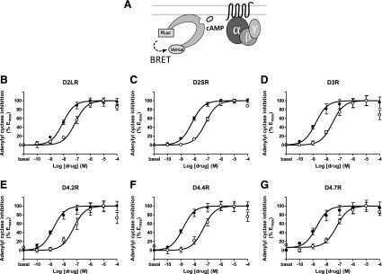
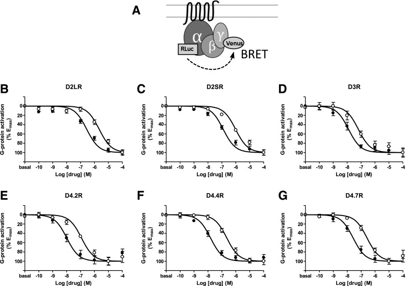
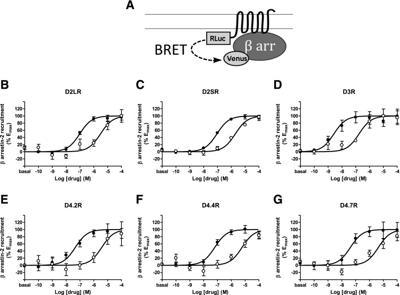
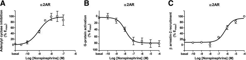
The Gαi/o-coupled dopamine D2-like receptor family comprises three subtypes: the D2 receptor (D2R), with short and long isoform variants (D2SR and D2LR), D3 receptor (D3R), and D4 receptor (D4R), with several polymorphic variants. The common overlap of norepinephrine innervation and D2-like receptor expression patterns prompts the question of a possible noncanonical action by norepinephrine. In fact, previous studies have suggested that norepinephrine can functionally interact with D4R. To our knowledge, significant interactions between norepinephrine and D2R or D3R receptors have not been demonstrated. By using radioligand binding and bioluminescent resonance energy transfer (BRET) assays in transfected cells, the present study attempted a careful comparison between dopamine and norepinephrine in their possible activation of all D2-like receptors, including the two D2R isoforms and the most common D4R polymorphic variants. Functional BRET assays included activation of G proteins with all Gαi/o subunits, adenylyl cyclase inhibition, and β arrestin recruitment. Norepinephrine acted as a potent agonist for all D2-like receptor subtypes, with the general rank order of potency of D3R > D4R ≥ D2SR ≥ D2L. However, for both dopamine and norepinephrine, differences depended on the Gαi/o protein subunit involved. The most striking differences were observed with Gαi2, where the rank order of potencies for both dopamine and norepinephrine were D4R > D2SR = D2LR >> D3R. Furthermore the results do not support the existence of differences in the ability of dopamine and norepinephrine to activate different human D4R variants. The potency of norepinephrine for adrenergic α2A receptor was only about 20-fold higher compared with D3R and D4R across the three functional assays.


Figures

Fig. 1.
Fig. 1.
(A) Scheme of the CAMYEL sensor configuration where the intramolecular conformational change triggered by cAMP results in a decrease in BRET due to RLuc8 and YFP changing their relative orientation from each other. (B–G) Dose–response experiments of adenylyl cyclase inhibition by dopamine (●) or norepinephrine (○) mediated by D2LR, D2SR, D3R, D4.2R, D4.4R, and D4.7R in HEK-293T cells transiently expressing the CAMYEL sensor and the indicated D2-like receptor: (B) D2LR, (C) D2SR, (D) D3R, (E) D4.2R, (F) D4.4R, or (G) D4.7R. Cells were treated with forskolin (10 µM) for 10 minutes followed by dopamine or norepinephrine and BRET values obtained by forskolin alone were subtracted from BRET values for each agonist concentration. Data represent the mean ± S.E.M. of 6 to 11 experiments performed in triplicate and are shown as a percentage of the maximal effect (see Table 2 for EC50 and Emax values and statistical comparisons).
Fig. 2.
Fig. 2.
(A) Scheme of the constructs used for G-protein activation BRET experiments where RLuc8 is fused to the Gαi1 subunit and mVenus is fused to the γ2 subunit. (B–G) Dose–response experiments of G protein activation by dopamine (●) or norepinephrine (○) mediated by D2LR, D2SR, D3R, D4.2R, D4.4R, and D4.7R in HEK-293T cells transiently expressing the G protein subunits Gαi1-RLuc8, unfused β1 and γ2-mVenus and the indicated D2-like receptor: (B) D2LR, (C) D2SR, (D) D3R, (E) D4.2R, (F) D4.4R, or (G) D4.7R. Cells were treated with coelenterazine H followed by increasing concentrations of dopamine or norepinephrine. After 10 minutes, BRET between G-protein subunits was measured as described in Materials and Methods. BRET values in the absence of ligands were subtracted from the BRET values for each agonist concentration. Data were fit by nonlinear regression to a sigmoidal dose–response relationship against the agonist concentration and are shown as a percentage of the maximal effect. Data represent the mean ± S.E.M. of 11 to 18 experiments performed in triplicate (see Table 3 for EC50 and Emax values and statistical comparisons).
Fig. 3.
Fig. 3.
(A) Scheme of the constructs used for β arrestin-2 recruitment-BRET experiments with different D2-like receptors fused to RLuc8 and β arrestin-2 fused to mVenus. (B–G) Dose–response experiments of β arrestin-2 recruitment by dopamine (●) or norepinephrine (○) mediated by (B) D2LR, (C) D2SR, (D) D3R, (E) D4.2R, (F) D4.4R, or (G) D4.7R in HEK-293T cells transiently expressing the indicated D2-like receptor fused to RLuc8, β arrestin-2-mVenus, and GRK2. Cells were treated with coelenterazine H followed by increasing concentrations of dopamine or norepinephrine. After 20 minutes, BRET was measured as described in Materials and Methods. BRET values in the absence of ligands were subtracted from the BRET values for each agonist concentration. Data were fit by nonlinear regression to a sigmoidal dose–response relationship against the agonist concentration and are shown as a percentage of the maximal effect. Data are the mean ± S.E.M. of 5 to 10 experiments performed in triplicate (see Table 4 for EC50 and Emax values and statistical comparisons).
Fig. 4.
Fig. 4.
(A–C) Dose–response experiments of norepinephrine on α2AR function. (A) Adenylyl cyclase inhibition assay using CAMYEL biosensor. (B) G protein activation assay using Gαi1 and γ2 subunits. (C) β Arrestin-2 recruitment assay using β arrestin-2-mVenus and GRK2. Conditions for each assay are identical to the ones described in Figures 1–3. Basal BRET values for forskolin alone (A) or in the absence of ligands (B, C) were subtracted from the BRET values for each agonist concentration. Data were fit by nonlinear regression to a sigmoidal dose–response relationship against the agonist concentration and are shown as a percentage of the maximal effect. Data are the mean ± S.E.M. of three to four experiments performed in triplicate (see Table 5 for EC50 and Emax values and statistical comparisons).

Similar articles

Cited by

References

    1. Agnati LF, Guidolin D, Guescini M, Genedani S, Fuxe K. (2010) Understanding wiring and volume transmission. Brain Res Brain Res Rev 64:137–159. - PubMed
    1. Antoni FA. (2012) New paradigms in cAMP signalling. Mol Cell Endocrinol 353:3–9. - PubMed
    1. Asghari V, Sanyal S, Buchwaldt S, Paterson A, Jovanovic V, Van Tol HHM. (1995) Modulation of intracellular cyclic AMP levels by different human dopamine D4 receptor variants. J Neurochem 65:1157–1165. - PubMed
    1. Atwood BK, Lopez J, Wager-Miller J, Mackie K, Straiker A. (2011) Expression of G protein-coupled receptors and related proteins in HEK293, AtT20, BV2, and N18 cell lines as revealed by microarray analysis. BMC Genomics 12:14. - PMC - PubMed
    1. Beaulieu J-M, Gainetdinov RR. (2011) The physiology, signaling, and pharmacology of dopamine receptors. Pharmacol Rev 63:182–217. - PubMed

Publication types

MeSH terms

LinkOut - more resources