. 2017 Feb 14;38(7):478-488.
doi: 10.1093/eurheartj/ehv760.

Coronary microcirculatory pathophysiology: can we afford it to remain a black box?

Affiliations

Coronary microcirculatory pathophysiology: can we afford it to remain a black box?

Axel R Pries et al. Eur Heart J. .

Abstract

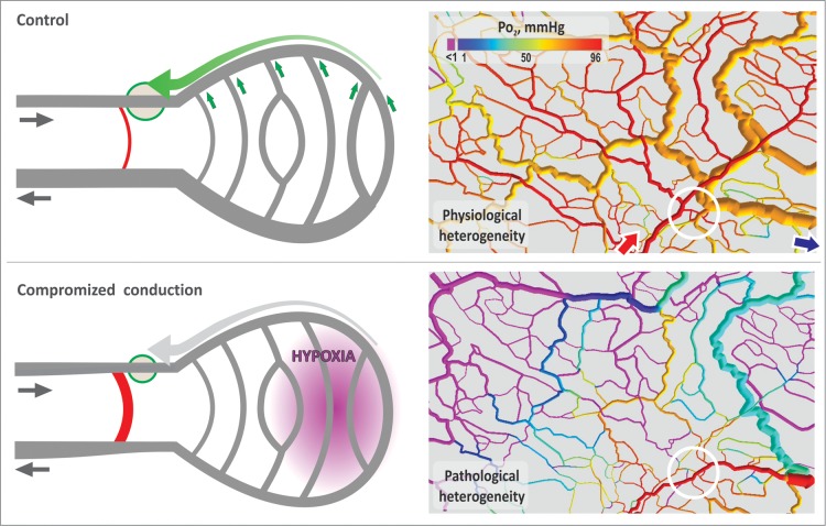
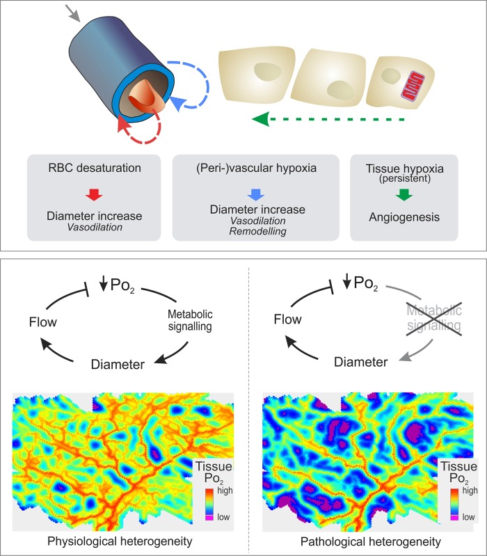
Coronary microvascular networks play the key role in determining blood flow distribution in the heart. Matching local blood supply to tissue metabolic demand entails continuous adaptation of coronary vessels via regulation of smooth muscle tone and structural dilated vessel diameter. The importance of coronary microcirculation for relevant pathological conditions including angina in patients with normal or near-normal coronary angiograms [microvascular angina (MVA)] and heart failure with preserved ejection fraction (HFpEF) is increasingly recognized. For MVA, clinical studies have shown a prevalence of up to 40% in patients with suspected coronary artery disease and a relevant impact on adverse cardiovascular events including cardiac death, stroke, and heart failure. Despite a continuously increasing number of corresponding clinical studies, the knowledge on pathophysiological cause-effect relations involving coronary microcirculation is, however, still very limited. A number of pathophysiological hypotheses for MVA and HFpEF have been suggested but are not established to a degree, which would allow definition of nosological entities, stratification of affected patients, or development of effective therapeutic strategies. This may be related to a steep decline in experimental (animal) pathophysiological studies in this area during the last 15 years. Since technology to experimentally investigate microvascular pathophysiology in the beating heart is increasingly, in principle, available, a concerted effort to build 'coronary microcirculatory observatories' to close this gap and to accelerate clinical progress in this area is suggested.

Keywords: CMVD; Conduction; Endothelial surface layer; Glycocalyx; HFpEF; MVA; Mathematical modelling; Metabolic regulation; Microvascular heterogeneity.


Figures

Figure 1
Figure 1
Model for vascular diameter adaptation in microvascular networks. Top: adaptation concept. Every vessel responds to local haemodynamic signals (wall shear stress, transmural pressure/circumferential wall stress), to metabolic signals, and to signals transmitted via upstream signal conduction through gap junctions in the vessel wall and downstream via convection of signal substances with the flowing blood. Bottom: modelling approach. In vivo experimental data from large microvascular networks are combined with theoretical simulations to derive a minimal set of adaptation rules. Network haemodynamics and oxygen distribution are calculated for all vessel segments. Segment adaptation of diameter and wall thickness in response to local conditions is determined according to an assumed set of adaptation rules. Haemodynamics and metabolics are re-calculated evoking further adaptation iteratively until convergence is achieved. Adaptation rules are optimized by comparing in vivo and calculated flow velocity for all vessels.,
Figure 2
Figure 2
Heterogeneity of microvascular networks. Computer reconstruction of a mesenteric microvascular network. White arrows indicate the main feeding (draining) arteriole (venule). The vessels are colour coded for wall shear stress (upper panel) and a metabolic signal (lower panel), which increases with decreasing pO2 as calculated by a computer model of vascular adaptation (see above)., Red arrows indicate two closely spaced capillaries with very different haemodynamic and metabolic conditions.
Figure 3
Figure 3
Consequences of compromised macro- and microvascular function. Coronary artery stenosis (left) and consequential increase of flow resistance lead to decreased bulk perfusion, global or regional supply-to-demand mismatch, and increased oxygen extraction (increased arteriovenous difference of oxygen content, AVD-O2). In the microcirculation, a generalized dysfunction on the arteriolar level may also lead to global tissue ischaemia. Typically, however, microvascular disturbance leads to an increased heterogeneity of flow and oxygen distribution (right). Underperfused tissue regions with low flow and high extraction may border to overperfused regions with high flow and low extraction through which blood is functionally shunted resulting in reduced mean arteriovenous difference of oxygen content.,
Figure 4
Figure 4
Conducted signals in vascular adaptation and effect of compromised conduction. Top: real vascular networks exhibit long and short arteriovenous flow pathways. Left: a short pathway (red) branches from an arteriole (green circle) that feeds a number of more distal capillaries. To prevent functional shunting, the feeding arteriole must exhibit a high diameter and low flow resistance relative to the short pathway. However, both vessels experience the same metabolic conditions, and the short connection exhibits high wall shear stress, which acts as growth signal. To prevent shunting, a strong compensating growth signal is required exclusively for the feeding arteriole. This signal is generated in response to metabolic conditions in the supported capillaries (small green arrows) and transmitted via connexins along the vessel wall to the feeding vessel (large green arrow). Thus, the flow in the feeding arteriole is maintained high and heterogeneity of oxygen distribution remains on an acceptable level. Right: a similar situation is shown for a network in the rat mesentery. The main arteriole (red arrow) is connected to the main draining venule of the network (blue arrow) via a short but narrow flow pathway (white circle). Bottom: if conduction is low or absent, the diameter of the feeding vessel relative to that of the short arteriovenous connection decreases (left panel and right panel, white circle). Thus, blood flow to distal regions is low, leading to a very heterogeneous oxygen distribution and large hypoxic areas.
Figure 5
Figure 5
Possible effects of compromised metabolic signalling in vascular adaptation. Top: metabolic signals for vascular diameter adaptation may be generated by red blood cells (red arrows), by the vessel wall and the perivascular region (blue arrows), and by parenchymal tissue cells (green arrows)., Bottom: tissue oxygen distribution in the presence of functional metabolic signalling (left) exhibiting a physiological level of heterogeneity and upon compromised metabolic signalling (right) with strongly increased spatial heterogeneity not compatible with normal tissue function.
Figure 6
Figure 6
Pathophysiological relations. Top: each coronary vessel, especially in the microcirculation, is subject to continuous adaptation to local conditions leading to changes in vessel phenotype and perfusion pattern (left). Deficits on the steps of this cascade correspond to different pathophysiological mechanisms (right). Bottom: for the pathophysiological mechanisms proposed here, hypothetical causal chains can be considered, which would link these mechanisms to the main conditions considered here, i.e. microvascular angina and heart failure with preserved ejection fraction.
Figure 7
Figure 7
Development of publication frequency in the area of coronary microcirculation. PubMed search: (((coronary microcirculation) OR (cardiac microcirculation) OR (myocardial microcirculation)) AND ‘humans’[MeSH Terms:noexp] NOT ‘animals’[MeSH Terms:noexp]); (((coronary microcirculation) OR (cardiac microcirculation) OR (myocardial microcirculation)) AND ‘animals’[MeSH Terms:noexp] NOT ‘humans’[MeSH Terms:noexp]) on 5 September 2015.
Figure 8
Figure 8
Intravital microscopy of the subepicardial microcirculation with standard and multi-photon microscopy (Langendorff preparation, rat heart; courtesy of H. Habazettl).

References

    1. Likoff W, Segal BL, Kasparian H. Paradox of normal selective coronary arteriograms in patients considered to have unmistakable coronary heart disease. N Engl J Med 1967;276:1063–1066. - PubMed
    1. Cannon RO III, Epstein SE. “Microvascular angina” as a cause of chest pain with angiographically normal coronary arteries. Am J Cardiol 1988;61:1338–1343. - PubMed
    1. Lanza GA, Crea F. Primary coronary microvascular dysfunction: clinical presentation, pathophysiology, and management. Circulation 2010;121:2317–2325. - PubMed
    1. Patel MR, Peterson ED, Dai D, Brennan JM, Redberg RF, Anderson HV, Brindis RG, Douglas PS. Low diagnostic yield of elective coronary angiography. N Engl J Med 2010;362:886–895. - PMC - PubMed
    1. Johnston N, Schenck-Gustafsson K, Lagerqvist B. Are we using cardiovascular medications and coronary angiography appropriately in men and women with chest pain? Eur Heart J 2011;32:1331–1336. - PubMed

MeSH terms