. 2016 Mar 1;86(9):800-7.
doi: 10.1212/WNL.0000000000002418. Epub 2016 Feb 3.

Neurofascin-155 IgG4 in chronic inflammatory demyelinating polyneuropathy

Affiliations

Neurofascin-155 IgG4 in chronic inflammatory demyelinating polyneuropathy

Jérôme J Devaux et al. Neurology. .

Abstract

Objective: We report the clinical and serologic features of Japanese patients with chronic inflammatory demyelinating polyneuropathy (CIDP) displaying anti-neurofascin-155 (NF155) immunoglobulin G4 (IgG4) antibodies.

Methods: In sera from 533 patients with CIDP, anti-NF155 IgG4 antibodies were detected by ELISA. Binding of IgG antibodies to central and peripheral nerves was tested.

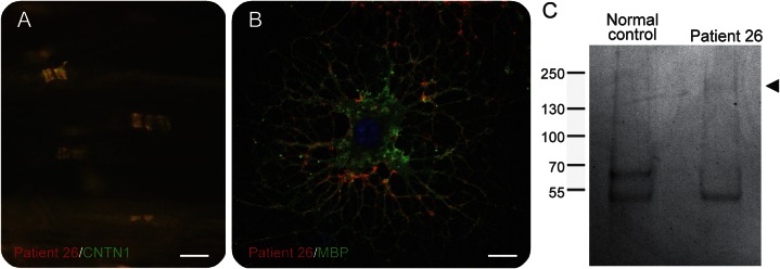
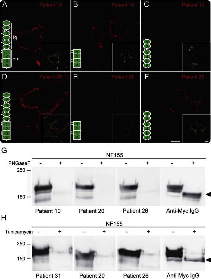
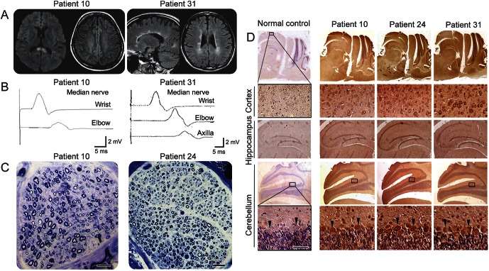
Results: Anti-NF155 IgG4 antibodies were identified in 38 patients (7%) with CIDP, but not in disease controls or normal participants. These patients were younger at onset as compared to 100 anti-NF155-negative patients with CIDP. Twenty-eight patients (74%) presented with sensory ataxia, 16 (42%) showed tremor, 5 (13%) presented with cerebellar ataxia associated with nystagmus, 3 (8%) had demyelinating lesions in the CNS, and 20 of 25 (80%) had poor response to IV immunoglobulin. The clinical features of the antibody-positive patients were statistically more frequent as compared to negative patients with CIDP (n = 100). Anti-NF155 IgG antibodies targeted similarly central and peripheral paranodes.

Conclusion: Anti-NF155 IgG4 antibodies were associated with a subgroup of patients with CIDP showing a younger age at onset, ataxia, tremor, CNS demyelination, and a poor response to IV immunoglobulin. The autoantibodies may serve as a biomarker to improve patients' diagnosis and guide treatments.


Figures

Figure 1
Figure 1. Glial NF155 is an autoantibody target in chronic inflammatory demyelinating polyneuropathy
(A) These are mouse sciatic nerve fibers immunostained for IgG antibodies (red) from a patient with chronic inflammatory demyelinating polyneuropathy (patient 26) and for CNTN1 (green) to stain the paranodes. Human IgG antibodies bound the paranodal regions and colocalized with CNTN1. (B) The IgG antibodies (red) from this patient recognized a glial antigen at the surface of oligodendrocytes in culture labeled for MBP (green). Scale bar = 10 μm. (C) The target antigens were immunoprecipitated from cultured oligodendrocytes, separated on sodium dodecyl sulfate–polyacrylamide gel electrophoresis gels, and stained with imperial blue. As control, IgG antibodies from a normal control (left) were used for immunoprecipitation. A protein band around 150–160 kDa (arrowhead) was specifically pulled down by the patient's IgG antibodies and identified as NF155 by mass spectrometry. Molecular weight markers are shown on the left in kilodalton. CNTN1 = contactin 1; IgG = immunoglobulin G; MBP = myelin basic protein; NF155 = neurofascin-155.
Figure 2
Figure 2. Anti-NF155 IgG antibodies target an N-glycosylated module
(A–F) Patients' IgG antibodies (red) were tested on living human embryonic kidney cells transfected with full-length Myc-tagged NF155 (A and D) or with constructs encoding solely the fibronectin type III (Fn) domains 1–4 (B and E) or the Ig domains 1–6 (C and F). A scheme of NF155 structure is inserted in each panel, showing the position of the putative N-glycosylation sites. Cells were then fixed and immunostained for Myc (green). Representative sera from patients with combined central and peripheral demyelination (patient 10) and chronic inflammatory demyelinating polyneuropathy (patient 25) are shown. The IgG antibodies from patient 10 (A–C) reacted against the Fn domains, whereas the IgG antibodies from patient 25 (D–F) recognize the Ig domains of NF155. The insets show the merge of the 2 channels (IgG and Myc). Scale bars = 10 μm. (G and H) Protein samples from NF155-transfected cells were untreated (−) or treated (+) with PNGaseF (G) or with tunicamycin (2 μg/mL; H). Proteins were then immunoblotted against Myc or representative sera from patients with chronic inflammatory demyelinating polyneuropathy (patients 10, 20, 26, and 31). The mouse anti-Myc antibodies recognized the N-glycosylated form of NF155 around 155 kDa and the unglycosylated protein core (arrowheads on the right). By contrast, patients' IgG antibodies only reacted against the N-glycosylated NF155. Molecular weight markers are shown on the left in kilodalton. IgG = immunoglobulin G; NF155 = neurofascin-155; PNGaseF = N-glycosidase F.
Figure 3
Figure 3. Laboratory findings of patients with combined central and peripheral demyelination
(A) Diffusion-weighted images in patient 10 showed signal abnormalities in the splenium of the corpus callosum. Fluid-attenuated inversion recovery images in patients 10 and 31 showed multiple sclerosis–like lesions in the juxtaventricular regions. (B) Median nerve motor conduction studies showed prolonged distal latencies and reduced conduction velocities with reduced amplitude and probable conduction block. (C) These are transverse sections of sural nerve biopsies stained with toluidine blue. Sural nerve biopsies revealed a moderate decrease in the number of large- and small-diameter fibers, some demyelinating changes, and axonal degeneration, but no cellular infiltration or onion-bulb formation. Scale bars = 50 μm. (D) The immunoreactivity of serum immunoglobulin G antibodies was examined in mouse cortex, hippocampus, and cerebellum, counterstained with hematoxylin. Note that the 3 sera strongly labeled Purkinje cells (arrowheads). Scale bar = 50 μm.

Comment in

Similar articles

Cited by

References

    1. Latov N. Diagnosis and treatment of chronic acquired demyelinating polyneuropathies. Nat Rev Neurol 2014;10:435–446. - PubMed
    1. Ng JK, Malotka J, Kawakami N, et al. Neurofascin as a target for autoantibodies in peripheral neuropathies. Neurology 2012;79:2241–2248. - PMC - PubMed
    1. Querol L, Nogales-Gadea G, Rojas-Garcia R, et al. Neurofascin IgG4 antibodies in CIDP associate with disabling tremor and poor response to IVIg. Neurology 2014;82:879–886. - PMC - PubMed
    1. Charles P, Tait S, Faivre-Sarrailh C, et al. Neurofascin is a glial receptor for the paranodin/Caspr-contactin axonal complex at the axoglial junction. Curr Biol 2002;12:217–220. - PubMed
    1. Bhat MA, Rios JC, Lu Y, et al. Axon-glia interactions and the domain organization of myelinated axons require neurexin IV/Caspr/paranodin. Neuron 2001;30:369–383. - PubMed

Publication types

MeSH terms

LinkOut - more resources