. 2016 Feb 3;36(5):1445-55.
doi: 10.1523/JNEUROSCI.0256-15.2016.

An In Vivo Pharmacological Screen Identifies Cholinergic Signaling as a Therapeutic Target in Glial-Based Nervous System Disease

Affiliations

An In Vivo Pharmacological Screen Identifies Cholinergic Signaling as a Therapeutic Target in Glial-Based Nervous System Disease

Liqun Wang et al. J Neurosci. .

Abstract

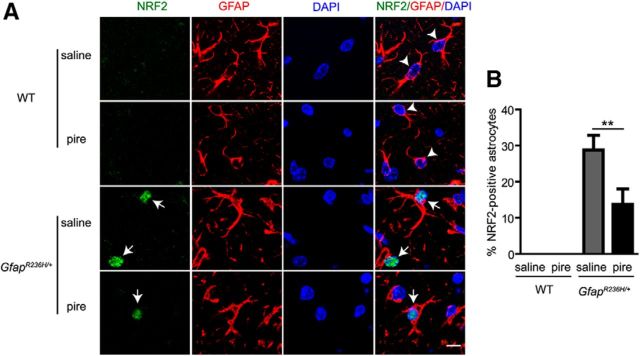
The role that glia play in neurological disease is poorly understood but increasingly acknowledged to be critical in a diverse group of disorders. Here we use a simple genetic model of Alexander disease, a progressive and severe human degenerative nervous system disease caused by a primary astroglial abnormality, to perform an in vivo screen of 1987 compounds, including many FDA-approved drugs and natural products. We identify four compounds capable of dose-dependent inhibition of nervous system toxicity. Focusing on one of these hits, glycopyrrolate, we confirm the role for muscarinic cholinergic signaling in pathogenesis using additional pharmacologic reagents and genetic approaches. We further demonstrate that muscarinic cholinergic signaling works through downstream Gαq to control oxidative stress and death of neurons and glia. Importantly, we document increased muscarinic cholinergic receptor expression in Alexander disease model mice and in postmortem brain tissue from Alexander disease patients, and that blocking muscarinic receptors in Alexander disease model mice reduces oxidative stress, emphasizing the translational significance of our findings. We have therefore identified glial muscarinic signaling as a potential therapeutic target in Alexander disease, and possibly in other gliopathic disorders as well.

Significance statement: Despite the urgent need for better treatments for neurological diseases, drug development for these devastating disorders has been challenging. The effectiveness of traditional large-scale in vitro screens may be limited by the lack of the appropriate molecular, cellular, and structural environment. Using a simple Drosophila model of Alexander disease, we performed a moderate throughput chemical screen of FDA-approved drugs and natural compounds, and found that reducing muscarinic cholinergic signaling ameliorated clinical symptoms and oxidative stress in Alexander disease model flies and mice. Our work demonstrates that small animal models are valuable screening tools for therapeutic compound identification in complex human diseases and that existing drugs can be a valuable resource for drug discovery given their known pharmacological and safety profiles.

Keywords: Alexander disease; Drosophila; chemical screen; cholinergic signaling; glia.


Figures

Figure 1.
Figure 1.
Screen design and results. Each compound from the Spectrum Collection was mixed with instant Drosophila medium individually at a final concentration of 100 μm. One-day-old Alexander disease model flies carrying a transgenic caspase reporter (repo-GAL4, UAS-GFAPR79H, UAS-CD8-PARP-Venus /+) were raised on drug-embedded food for a total of 10 d, with transfer to a vial with fresh drug-embedded food every 3 d. Caspase activation was then monitored by determining the number of cleaved PARP-positive cells in the brain using immunohistochemical (IHC) detection on sections from paraffin-embedded material.
Figure 2.
Figure 2.
Glycopyrrolate reduces GFAP toxicity in Alexander disease model flies. A, Double-label immunofluorescence reveals both glial (top panel, arrows) and neuronal (bottom panel, arrows) cell death in Alexander disease model flies. Apoptotic cells are labeled by TUNEL. Repo is a glial cell marker, and elav is a neuronal cell marker. DAPI labels nuclei. Scale bar, 3 μm. Genotype: repo-GAL4, UAS-GFAPR79H/+. Flies were 15-d-old. B, Glycopyrrolate significantly reduces the total number of TUNEL-positive cells at 50 μm. ***p < 0.001 (two-tailed t test). n ≥ 6 per concentration. Flies were 15-d-old and were treated with drug for 15 d. Genotype: repo-GAL4, UAS-GFAPR79H/+. Control flies were treated with solvent (water) only. Arrows indicate TUNEL-positive cells. Scale bar, 20 μm. C, Glycopyrrolate significantly reduces the percentage of flies with seizures at 50 μm. **p < 0.01 (χ2 test). n > 100 per concentration. Flies were 3-d-old and were treated with drug for 3 d. Genotype: repo-GAL4, UAS-GFAPR79H/+. Control (ctrl) flies were treated with solvent (water) only. The schematic represents the method for seizure induction.
Figure 3.
Figure 3.
Compounds inhibiting mAChR reduce GFAP toxicity in Alexander disease model flies. A–C, Dose–response analysis of three compounds targeting mAChR. mAChR antagonists, atropine (A) and scopolamine (B) reduce the number of cleaved PARP-positive cells in a dose-dependent manner, whereas the agonist, pilocarpine (C), increases the number of cleaved PARP-positive cells in a dose-dependent manner. *p < 0.05 (one-way ANOVA with Tukey's multiple-comparison test). **p < 0.01 (one-way ANOVA with Tukey's multiple-comparison test). n ≥ 6 per concentration. Flies were 10-d-old and were treated with drugs for 10 d. Genotype: repo-GAL4, UAS-GFAPR79H, UAS-CD8-PARP-Venus/+. D–F, Cell death analysis in mAChR compound-fed flies. Atropine (D) and scopolamine (E) significantly reduce, whereas pilocarpine (F) increases, the number of TUNEL-positive cells in Alexander disease model flies. *p < 0.05 (two-tailed t test). **p < 0.01 (two-tailed t test). n ≥ 6 per concentration. Flies were 15-d-old and were treated with drugs for 15 d. Genotype: repo-GAL4, UAS-GFAPR79H/+. Ctrl: solvent (water)-fed flies. Arrows indicate TUNEL-positive cells. Scale bar, 20 μm. G–I, Seizure analysis in mAChR compound-fed flies. Atropine (G) and scopolamine (H) reduce, whereas pilocarpine (I) increases, the percentage of flies with seizures. **p < 0.01 (χ2 test). ***p < 0.001 (χ2 test). n > 100 per concentration. Flies were 3-d-old and were treated with drugs for 3 d. Genotype: repo-GAL4, UAS-GFAPR79H/+. J, Western blot shows equivalent GFAP protein levels in control (ctrl) and drug-fed flies. Flies were 3-d-old and were treated with drug for 3 d. The blot was reprobed with an antibody for actin to illustrate equivalent protein loading. Genotype: repo-GAL4, UAS-GFAPR79H/+. Control (Ctrl) flies were treated with solvent (water) only.
Figure 4.
Figure 4.
mAChR compounds modulate oxidative stress in Alexander disease model flies. A, Double-label immunofluorescence shows activation of the oxidative stress reporter, GstD1-lacZ, in glial cells (arrows) of Alexander disease model flies. Repo marks glia, and elav marks neurons. DAPI labels nuclei. Scale bar, 3 μm. Genotype: GstD1-lacZ/+; repo-GAL4, UAS-GFAPR79H/+. Flies were 15-d-old. B–D, Oxidative stress analysis in mAChR compound-fed flies. Atropine (B) and scopolamine (C) reduce, whereas pilocarpine (D) increases, the number of β-galactosidase-positive cells in Alexander disease model flies. *p < 0.05 (two-tailed t test). ***p < 0.001 (two-tailed t test). n ≥ 6 per concentration. Flies were 15-d-old and were treated with drugs for 15 d. Genotype: GstD1-lacZ/+; repo-GAL4, UAS-GFAPR79H/+. Arrows indicate β-galactosidase-positive cells. Scale bar, 20 μm.
Figure 5.
Figure 5.
Genetic inhibition of mAChR pathway reduces GFAP toxicity in Alexander disease model flies. In all panels, GFAP-R79H indicates repo-GAL4, UAS-GFAPR79H/+. mAChR-A RNAi indicates UAS-mAChR-A RNAi (A–E), and Gαq RNAi indicates UAS-Gαq RNAi (F–J). A, F, Reducing expression of mAChR-A or Gαq with transgenic RNAi significantly reduces the number of TUNEL-positive cells. **p < 0.01. ***p < 0.001. n ≥ 6 per genotype. Control (+): repo-GAL4, UAS-GFAPR79H/+. B, G, Reducing expression of mAChR-A or Gαq significantly decreases the percentage of flies with seizures. **p < 0.01. ***p < 0.001. n > 100 per genotype. Control (+): repo-GAL4, UAS-GFAPR79H/+. C, H, Decreased expression of mAChR-A or Gαq reduces the number of β-galactosidase-positive cells in Alexander disease model flies. *p < 0.05. ***p < 0.001. n ≥ 6 per genotype. Control (+): GstD1-lacZ/+; repo-GAL4, UAS-GFAPR79H/+. D, I, Western blots demonstrate equivalent GFAP levels in GFAPR79H transgenic flies alone and in combination with mAChR-A RNAi or Gαq RNAi lines. The blots were reprobed with an antibody to actin to illustrate equivalent protein loading. Control (+): repo-GAL4, UAS-GFAPR79H/+. E, J, RT-PCR shows reduced mAChR-A and Gαq mRNA levels in transgenic RNAi lines, using da-GAL4 or repo-GAL4 as drivers. Rpl32 was used as a control. ***p < 0.001. n = 3 biological replicates. Control (+): da-GAL4/+ (E) and repo-GAL4/+ (J). A, C, F, H, Flies were 20-d-old. B, D, E, G, I, J, Flies were 1-d-old. Statistical tests: A, E, F, J, one-way ANOVA with Tukey's multiple-comparison test; B, G, χ2 test; C, H, two-tailed t test.
Figure 6.
Figure 6.
Increased mAChRs in astrocytes of Alexander disease model mice. A, Double-label immunofluorescence reveals increased immunostaining with pan-mAChR antibody, M35, in the astrocytes of 6-month-old GFAPR239H/+ mice (arrows). GFAP is an astrocytic marker. Asterisks show blood vessels. DAPI labels nuclei. Scale bar, 20 μm. B, Double-label immunofluorescence reveals upregulation of the M1 receptor in astrocytes of 6-month-old GFAPR239H/+ mice (arrows). DAPI labels nuclei. Scale bar, 10 μm. C, Western blot demonstrates increased expression of M1 receptor in the corpus callosum of 6-month-old GFAPR236H/+ mice. The blot was reprobed with an antibody to GAPDH to illustrate equivalent protein loading. n = 4 biological replicates. *p < 0.05 (two-tailed Student's t test).
Figure 7.
Figure 7.
Increased expression of M1 receptor in Alexander disease patients. A, Double-label immunofluorescence reveals upregulation of M1 receptor in the astrocytes of an Alexander disease patient (top and middle panels, arrows). Arrowheads indicate Rosenthal fibers. GFAP marks astrocytes. DAPI labels nuclei. Scale bar, 10 μm. B, Western blot shows significantly increased expression of M1 receptors in the white matter of Alexander disease patients. The blot was reprobed with an antibody to GAPDH to illustrate equivalent protein loading. *p < 0.05 (two-tailed Student's t test).
Figure 8.
Figure 8.
Pirenzepine treatment reduces NRF2 in Alexander disease model mice. A, Double-label immunofluorescence reveals expression of NRF2 in astrocytes of Alexander disease model mice (bottom two rows), but not in age-matched wild-type control mice (top two rows). GFAP marks astrocytes. DAPI labels nuclei. Arrows indicate NRF2-positive astrocytes. Arrowheads indicate NRF2-negative astrocytes. Scale bar, 10 μm. B, Quantification in the hippocampal CA1 region shows a significant reduction of the percentage of NRF2-positive astrocytes in pirenzepine (pire)-treated Alexander disease model mice compared with saline-treated Alexander disease mice. **p < 0.01 (one-way ANOVA with Tukey's multiple-comparison test). Fifty astrocytes per animal and 5 or 6 animals per genotype/treatment were counted. Mice were 8 weeks old and were treated with 20 mg/kg pirenzepine or saline for 10 d.

Similar articles

Cited by

References

    1. Agrawal T, Sadaf S, Hasan G. Genetic RNAi screen for IP3/Ca2+ coupled GPCRs in Drosophila identifies the PdfR as a regulator of insect flight. PLoS Genet. 2013;9:e1003849. doi: 10.1371/journal.pgen.1003849. - DOI - PMC - PubMed
    1. Anisuzzaman AS, Uwada J, Masuoka T, Yoshiki H, Nishio M, Ikegaya Y, Takahashi N, Matsuki N, Fujibayashi Y, Yonekura Y, Momiyama T, Muramatsu I. Novel contribution of cell surface and intracellular M1-muscarinic acetylcholine receptors to synaptic plasticity in hippocampus. J Neurochem. 2013;126:360–371. doi: 10.1111/jnc.12306. - DOI - PubMed
    1. Bonini NM, Fortini ME. Human neurodegenerative disease modeling using Drosophila. Annu Rev Neurosci. 2003;26:627–656. doi: 10.1146/annurev.neuro.26.041002.131425. - DOI - PubMed
    1. Brand AH, Perrimon N. Targeted gene expression as a means of altering cell fates and generating dominant phenotypes. Development. 1993;118:401–415. - PubMed
    1. Brenner M, Johnson AB, Boespflug-Tanguy O, Rodriguez D, Goldman JE, Messing A. Mutations in GFAP, encoding glial fibrillary acidic protein, are associated with Alexander disease. Nat Genet. 2001;27:117–120. doi: 10.1038/83679. - DOI - PubMed

Publication types

MeSH terms

LinkOut - more resources