

Left Superior Temporal Gyrus Is Coupled to Attended Speech in a Cocktail-Party Auditory Scene
- PMID: 26843641
- PMCID: PMC6601992
- DOI: 10.1523/JNEUROSCI.1730-15.2016
Left Superior Temporal Gyrus Is Coupled to Attended Speech in a Cocktail-Party Auditory Scene
Abstract
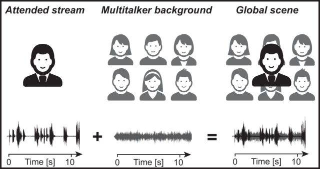
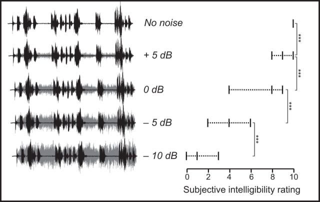
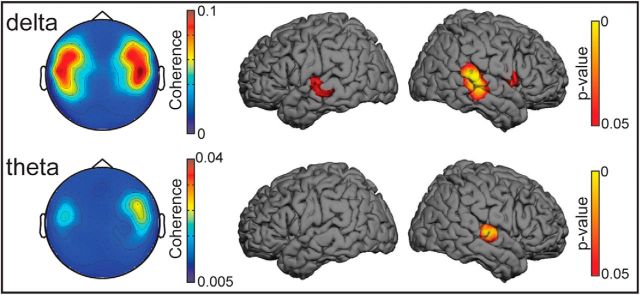
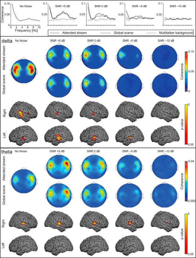
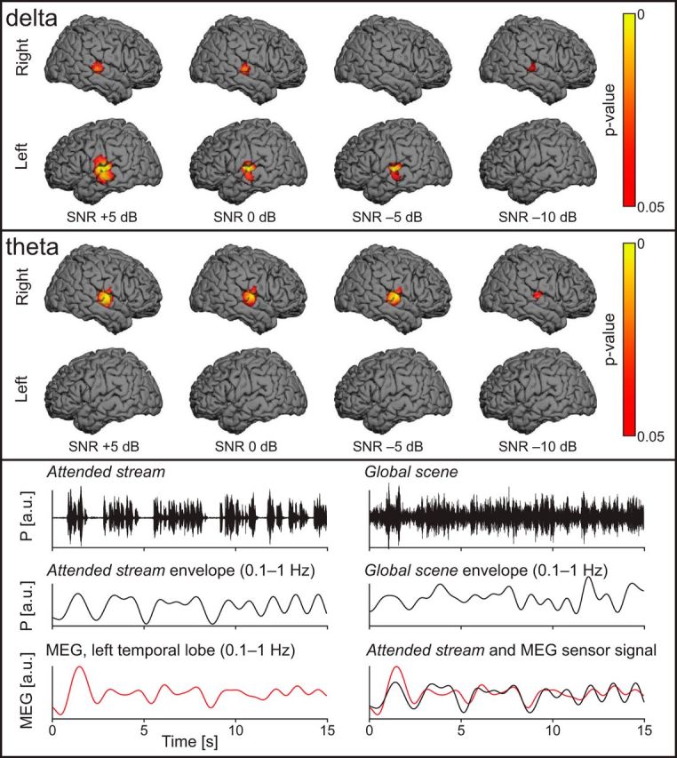
Using a continuous listening task, we evaluated the coupling between the listener's cortical activity and the temporal envelopes of different sounds in a multitalker auditory scene using magnetoencephalography and corticovocal coherence analysis. Neuromagnetic signals were recorded from 20 right-handed healthy adult humans who listened to five different recorded stories (attended speech streams), one without any multitalker background (No noise) and four mixed with a "cocktail party" multitalker background noise at four signal-to-noise ratios (5, 0, -5, and -10 dB) to produce speech-in-noise mixtures, here referred to as Global scene. Coherence analysis revealed that the modulations of the attended speech stream, presented without multitalker background, were coupled at ∼0.5 Hz to the activity of both superior temporal gyri, whereas the modulations at 4-8 Hz were coupled to the activity of the right supratemporal auditory cortex. In cocktail party conditions, with the multitalker background noise, the coupling was at both frequencies stronger for the attended speech stream than for the unattended Multitalker background. The coupling strengths decreased as the Multitalker background increased. During the cocktail party conditions, the ∼0.5 Hz coupling became left-hemisphere dominant, compared with bilateral coupling without the multitalker background, whereas the 4-8 Hz coupling remained right-hemisphere lateralized in both conditions. The brain activity was not coupled to the multitalker background or to its individual talkers. The results highlight the key role of listener's left superior temporal gyri in extracting the slow ∼0.5 Hz modulations, likely reflecting the attended speech stream within a multitalker auditory scene.
Significance statement: When people listen to one person in a "cocktail party," their auditory cortex mainly follows the attended speech stream rather than the entire auditory scene. However, how the brain extracts the attended speech stream from the whole auditory scene and how increasing background noise corrupts this process is still debated. In this magnetoencephalography study, subjects had to attend a speech stream with or without multitalker background noise. Results argue for frequency-dependent cortical tracking mechanisms for the attended speech stream. The left superior temporal gyrus tracked the ∼0.5 Hz modulations of the attended speech stream only when the speech was embedded in multitalker background, whereas the right supratemporal auditory cortex tracked 4-8 Hz modulations during both noiseless and cocktail-party conditions.
Keywords: coherence analysis; magnetoencephalography; speech in noise.
Copyright © 2016 the authors 0270-6474/16/361597-11$15.00/0.
Figures
Similar articles
-
Cortical Tracking of Speech-in-Noise Develops from Childhood to Adulthood.J Neurosci. 2019 Apr 10;39(15):2938-2950. doi: 10.1523/JNEUROSCI.1732-18.2019. Epub 2019 Feb 11. J Neurosci. 2019. PMID: 30745419 Free PMC article.
-
Cortical Representations of Speech in a Multitalker Auditory Scene.J Neurosci. 2017 Sep 20;37(38):9189-9196. doi: 10.1523/JNEUROSCI.0938-17.2017. Epub 2017 Aug 18. J Neurosci. 2017. PMID: 28821680 Free PMC article.
-
The Right Temporoparietal Junction Supports Speech Tracking During Selective Listening: Evidence from Concurrent EEG-fMRI.J Neurosci. 2017 Nov 22;37(47):11505-11516. doi: 10.1523/JNEUROSCI.1007-17.2017. Epub 2017 Oct 23. J Neurosci. 2017. PMID: 29061698 Free PMC article.
-
Stimulus-dependent activations and attention-related modulations in the auditory cortex: a meta-analysis of fMRI studies.Hear Res. 2014 Jan;307:29-41. doi: 10.1016/j.heares.2013.08.001. Epub 2013 Aug 11. Hear Res. 2014. PMID: 23938208 Review.
-
[Auditory perception and language: functional imaging of speech sensitive auditory cortex].Rev Neurol (Paris). 2001 Sep;157(8-9 Pt 1):837-46. Rev Neurol (Paris). 2001. PMID: 11677406 Review. French.
Cited by
-
Slow phase-locked modulations support selective attention to sound.Neuroimage. 2022 May 15;252:119024. doi: 10.1016/j.neuroimage.2022.119024. Epub 2022 Feb 26. Neuroimage. 2022. PMID: 35231629 Free PMC article.
-
Cortical Tracking of Speech-in-Noise Develops from Childhood to Adulthood.J Neurosci. 2019 Apr 10;39(15):2938-2950. doi: 10.1523/JNEUROSCI.1732-18.2019. Epub 2019 Feb 11. J Neurosci. 2019. PMID: 30745419 Free PMC article.
-
The functional and structural characteristics of the emotion network in alexithymia.Neuropsychiatr Dis Treat. 2018 Apr 12;14:991-998. doi: 10.2147/NDT.S154601. eCollection 2018. Neuropsychiatr Dis Treat. 2018. PMID: 29695908 Free PMC article.
-
Decreased inter-hemispheric interactions but increased intra-hemispheric integration during typical aging.Aging (Albany NY). 2019 Nov 21;11(22):10100-10115. doi: 10.18632/aging.102421. Epub 2019 Nov 21. Aging (Albany NY). 2019. PMID: 31761785 Free PMC article.
-
Aberrant Changes in Cortical Complexity in Right-Onset Versus Left-Onset Parkinson's Disease in Early-Stage.Front Aging Neurosci. 2021 Nov 8;13:749606. doi: 10.3389/fnagi.2021.749606. eCollection 2021. Front Aging Neurosci. 2021. PMID: 34819848 Free PMC article.
References
Publication types
MeSH terms
LinkOut - more resources
Full Text Sources
Other Literature Sources
Miscellaneous