. 2016 Feb 3;36(5):1660-8.
doi: 10.1523/JNEUROSCI.0895-15.2016.

Persistent Electrical Activity in Primary Nociceptors after Spinal Cord Injury Is Maintained by Scaffolded Adenylyl Cyclase and Protein Kinase A and Is Associated with Altered Adenylyl Cyclase Regulation

Affiliations

Persistent Electrical Activity in Primary Nociceptors after Spinal Cord Injury Is Maintained by Scaffolded Adenylyl Cyclase and Protein Kinase A and Is Associated with Altered Adenylyl Cyclase Regulation

Alexis Bavencoffe et al. J Neurosci. .

Abstract

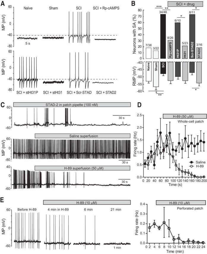
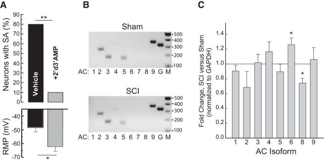
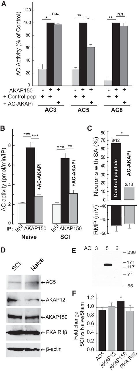
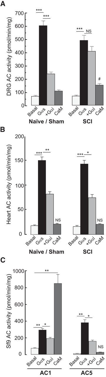
Little is known about intracellular signaling mechanisms that persistently excite neurons in pain pathways. Persistent spontaneous activity (SA) generated in the cell bodies of primary nociceptors within dorsal root ganglia (DRG) has been found to make major contributions to chronic pain in a rat model of spinal cord injury (SCI) (Bedi et al., 2010; Yang et al., 2014). The occurrence of SCI-induced SA in a large fraction of DRG neurons and the persistence of this SA long after dissociation of the neurons provide an opportunity to define intrinsic cell signaling mechanisms that chronically drive SA in pain pathways. The present study demonstrates that SCI-induced SA requires continuing activity of adenylyl cyclase (AC) and cAMP-dependent protein kinase (PKA), as well as a scaffolded complex containing AC5/6, A-kinase anchoring protein 150 (AKAP150), and PKA. SCI caused a small but significant increase in the expression of AKAP150 but not other AKAPs. DRG membranes isolated from SCI animals revealed a novel alteration in the regulation of AC. AC activity stimulated by Ca(2+)-calmodulin increased, while the inhibition of AC activity by Gαi showed an unexpected and dramatic decrease after SCI. Localized enhancement of the activity of AC within scaffolded complexes containing PKA is likely to contribute to chronic pathophysiological consequences of SCI, including pain, that are promoted by persistent hyperactivity in DRG neurons.

Significance statement: Chronic neuropathic pain is a major clinical problem with poorly understood mechanisms and inadequate treatments. Recent findings indicate that chronic pain in a rat SCI model depends upon hyperactivity in dorsal root ganglia (DRG) neurons. Although cAMP signaling is involved in many forms of neural plasticity, including hypersensitivity of nociceptors in the presence of inflammatory mediators, our finding that continuing cAMP-PKA signaling is required for persistent SA months after SCI and long after isolation of nociceptors is surprising. The dependence of ongoing SA upon AKAP150 and AC5/6 was unknown. The discovery of a dramatic decrease in Gαi inhibition of AC activity after SCI is novel for any physiological system and potentially has broad implications for understanding chronic pain mechanisms.

Keywords: A-kinase anchoring protein; DRG; cAMP; chronic pain; hyperexcitability; spontaneous activity.


Figures

Figure 1.
Figure 1.
Anchored PKA activity maintains SCI-induced SA in dissociated small L4/L5 DRG neurons. A, Examples of SA and RMP after the indicated treatments. Dashed line indicates least negative acceptable RMP (−40 mV) for sampled neurons. B, Attenuation of SCI-induced SA and depolarized RMP by inhibitors of PKA and AKAP function. The ratio above each bar denotes the number of neurons with SA/the number of neurons sampled. Statistical comparisons of SA incidence were made with Fisher's exact tests on the indicated pairs of groups (paired experiments were run on the same batches of neurons in different coverslips on the same day). Comparisons of RMP (mean ± SEM) were made with one-way ANOVA followed by Bonferroni post hoc tests: *p < 0.05; **p < 0.01; ***p < 0.001. C, Examples of ongoing SA in dissociated DRG neurons and its rapid reduction by inhibitors of PKA and AKAP function. Top, Cell recorded under whole-cell configuration with a higher resistance patch pipette containing 100 nm STAD-2 in pipette solution. Middle, Cell recorded with lower resistance patch pipette and superfused with saline (same as bath solution). Bottom, Cell recorded with a lower resistance pipette and superfused with H-89 in bath solution. D, Time course of the effects of superfused 50 μm H-89 on SA firing rate (mean ± SEM). Two-way ANOVA revealed significant effects of H-89 treatment (n = 6 neurons) versus controls (n = 10) and of time after treatment. E, Prolonged recordings of dissociated DRG neurons with the perforated patch method shows that 10 μm H-89 eventually suppresses ongoing SA. Left, Examples of SA recorded 1 min before and at the indicated times after superfusion of 10 μm H-89. Right, Time course of the effects of 10 μm H-89 treatment on mean SA firing rate recorded with perforated patch. Each point represents the mean ± SEM (n = 3 cells) of the average spike frequency in each cell during the indicated 2 min period. MP, Membrane potential (includes action potentials).
Figure 2.
Figure 2.
AC activity maintains SCI-induced SA. A, Attenuation of SCI-induced SA and depolarized RMP by an AC inhibitor (2′deoxy-3′AMP). The proportion of neurons exhibiting SA was analyzed with Fisher's exact test (**p < 0.01; 10 cells per group); RMP analyzed with unpaired t test (*p < 0.05). B, Agarose gel of RT-PCR products from RNA isolated from Sham and SCI rats of AC isoforms and GAPDH control after 40 cycles. Primers used are indicated in Table 1. C, qRT-PCR of AC isoform expression in DRGs from Sham and SCI rats. Fold change (2ΔΔCt) after SCI ± SD is plotted. All samples were normalized to GAPDH controls (n = 3, performed in triplicate). For description of statistical analysis, see Materials and Methods.
Figure 3.
Figure 3.
Disruption of AKAP150-anchored AC in DRGs by an AC5/6-AKAP150-selective disrupting peptide (AC-AKAPi) attenuates SCI-induced SA. A, AC-AKAPi is selective for AC5 and does not disrupt association of AC3 or AC8 with AKAP150. HEK293 cells were transfected with AKAP150 and the indicated AC isoforms. Samples were immunoprecipitated with anti-AKAP150 in the presence of control or disrupting peptides (10 μm) during cell lysis, and the associated stimulated AC activity was measured (mean ± SEM, n = 3 performed in duplicate, normalized to control peptide with AKAP150). *p < 0.05 (paired t test on non-normalized data). **p < 0.01 (paired t test on non-normalized data). B, AKAP150-associated AC activity is reduced by AC-AKAPi in DRGs from sham and SCI rats. Samples were immunoprecipitated with control IgG or anti-AKAP150 in the presence or absence of AC-AKAPi (20 μm), and associated AC activity was measured (mean ± SEM, n = 3). ***p < 0.001 (unpaired t test). C, Attenuation of SCI-induced SA by AC-AKAPi. *p < 0.05 (Fisher's exact test). D, F, AKAP150, but not AC5, protein expression in DRGs is slightly enhanced by SCI (mean ± SEM, n = 3–5). *p < 0.05 (paired t test of SCI vs Sham controls). E, Characterization of anti-AC5. WB of membranes from Sf9 cells expressing AC3, AC5, or AC6. n.s., Not significant.
Figure 4.
Figure 4.
SCI reduces Gαi inhibition of AC activity in DRG membranes and increases Ca2+/calmodulin-stimulated activity without changing basal or total Gαs-stimulated AC activity. AC activity was measured in membranes prepared from (A) DRGs (n = 4), (B) heart (n = 3), or (C) Sf9 cells expressing AC1 or AC5 (n = 3) under basal conditions or in the presence of Gαs (100 nm), Gαs plus Gαi (1 μm), or Ca2+/calmodulin (CaM; 100 μm/300 nm). *p < 0.05 (unpaired t tests). **p < 0.01 (unpaired t tests). ***p < 0.001 (unpaired t tests). #Significant difference in CaM-stimulated AC responses of SCI versus Naive/Sham animals. NS, Not significant.

References

    1. Aley KO, Levine JD. Role of protein kinase A in the maintenance of inflammatory pain. J Neurosci. 1999;19:2181–2186. - PMC - PubMed
    1. Araldi D, Ferrari LF, Levine JD. Repeated mu-opioid exposure induces a novel form of the hyperalgesic priming model for transition to chronic pain. J Neurosci. 2015a;35:12502–12517. doi: 10.1523/JNEUROSCI.1673-15.2015. - DOI - PMC - PubMed
    1. Araldi D, Ferrari LF, Levine JD. Adenosine-A1 receptor agonist induced hyperalgesic priming type II. Pain. 2015b Advance online publication. Retrieved Nov. 14, 2015. - PMC - PubMed
    1. Basso DM, Beattie MS, Bresnahan JC. A sensitive and reliable locomotor rating scale for open field testing in rats. J Neurotrauma. 1995;12:1–21. doi: 10.1089/neu.1995.12.1. - DOI - PubMed
    1. Bedi SS, Yang Q, Crook RJ, Du J, Wu Z, Fishman HM, Grill RJ, Carlton SM, Walters ET. Chronic spontaneous activity generated in the somata of primary nociceptors is associated with pain-related behavior after spinal cord injury. J Neurosci. 2010;30:14870–14882. doi: 10.1523/JNEUROSCI.2428-10.2010. - DOI - PMC - PubMed

Publication types

Substances