. 2016 Feb 3;36(5):1669-81.
doi: 10.1523/JNEUROSCI.1928-15.2016.

Sex Differences in GABAA Signaling in the Periaqueductal Gray Induced by Persistent Inflammation

Affiliations

Sex Differences in GABAA Signaling in the Periaqueductal Gray Induced by Persistent Inflammation

Karen J Tonsfeldt et al. J Neurosci. .

Abstract

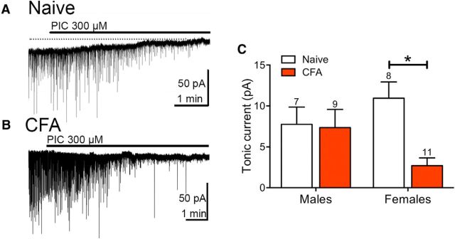
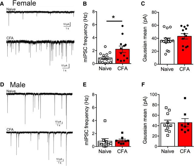
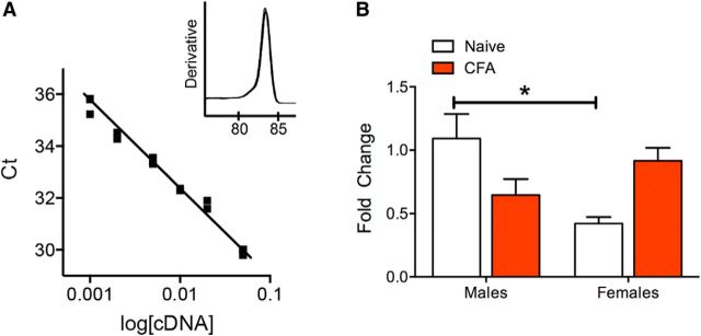
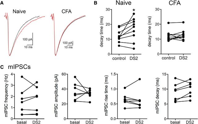
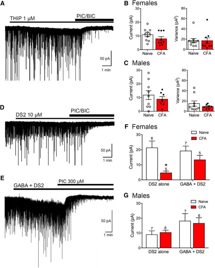
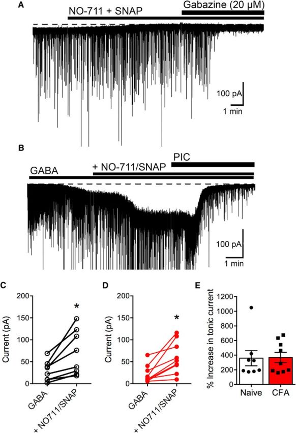
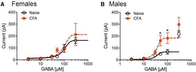
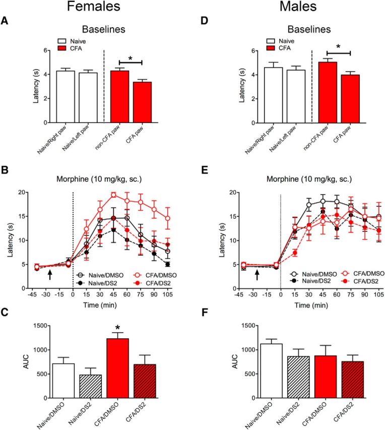
The ventrolateral periaqueductal gray (vlPAG) is a key structure in the descending pain modulatory circuit. Activation of the circuit occurs via disinhibition of GABAergic inputs onto vlPAG output neurons. In these studies, we tested the hypothesis that GABAergic inhibition is increased during persistent inflammation, dampening activation of the descending circuit from the vlPAG. Our results indicate that persistent inflammation induced by Complete Freund's adjuvant (CFA) modulates GABA signaling differently in male and female rats. CFA treatment results in increased presynaptic GABA release but decreased high-affinity tonic GABAA currents in female vlPAG neurons. These effects are not observed in males. The tonic currents in the vlPAG are dependent on GABA transporter activity and are modulated by agonists that activate GABAA receptors containing the δ subunit. The GABAA δ agonist THIP (gaboxadol) induced similar amplitude currents in naive and CFA-treated rats. In addition, a positive allosteric modulator of the GABAA δ subunit, DS2 (4-chloro-N-[2-(2-thienyl)imidazo[1,2-a]pyridin-3-yl]benzamide), increased tonic currents. These results indicate that GABAA δ receptors remain on the cell surface but are less active in CFA-treated female rats. In vivo behavior studies showed that morphine induced greater antinociception in CFA-treated females that was reversed with microinjections of DS2 directly into the vlPAG. DS2 did not affect morphine antinociception in naive or CFA-treated male rats. Together, these data indicate that sex-specific adaptations in GABAA receptor signaling modulate opioid analgesia in persistent inflammation. Antagonists of GABAA δ receptors may be a viable strategy for reducing pain associated with persistent inflammation, particularly in females.

Significance statement: These studies demonstrate that GABA signaling is modulated in the ventrolateral periaqueductal gray by persistent inflammation differently in female and male rats. Our results indicate that antagonists or negative allosteric modulators of GABAA δ receptors may be an effective strategy to alleviate chronic inflammatory pain and promote opioid antinociception, especially in females.

Keywords: GABAA; chronic pain; descending pain control; opioid; sex difference; tonic current.


Figures

Figure 1.
Figure 1.
GABAA-mediated tonic currents are reduced in CFA-treated female rats. A, The selective GABAA antagonist picrotoxin (PIC) induced outward deflection of the holding current at −70 mV and inhibited sIPSCs in naive rats. B, The deflection in holding current at −70 mV was reduced in a female rat pretreated with CFA 4–7 d before recording. C, Tonic currents were not affected in male rats but were reduced in female rats following CFA treatment (two-way ANOVA, effect of treatment; F(1,31) = 5.68, p = 0.024; Sidak's multiple-comparison test, *p < 0.05).
Figure 2.
Figure 2.
Persistent inflammation increases GABA release in female rats. A, Representative traces of GABAergic mIPSCs in naive (top) and CFA-treated (bottom) female PAG neurons. B, The frequency of mIPSCs is increased in CFA-treated compared with female rats (Mann–Whitney U = 40, *p = 0.02). C, The mean amplitude of mIPSCs determined from a Gaussian fit to the amplitude histogram was not altered in CFA-treated female rats (t(24) = 1.15, p = 0.26). D, Representative traces of GABAergic mIPSCs in naive (top) and CFA-treated (bottom) male PAG neurons. E, The mIPSC frequency in male rats was not altered by CFA treatment (Mann–Whitney U = 19, p = 0.11). F, Mean amplitude of mIPSCs was also not altered in male rats pretreated with CFA (t(16) = 0.096, p = 0.92).
Figure 3.
Figure 3.
GABAA δ mRNA expression from vlPAG are lower in female rats. A, The designed GABAA δ primer efficiency was 100% (slope: −3.36, r2 = 0.99) and produced a single product (inset). B, There was a significant interaction between treatment and sex (two-way ANOVA, F(3,17) = 3.38, p = 0.0093) when comparing relative GABAA δ mRNA expression. Naive female rats had lower levels than naive male rats (Tukey's post hoc, *p < 0.05). Although there was a strong trend toward an increase in expression with CFA treatment in female rats and a decrease in expression with CFA treatment in males, neither reached significance (N = 4–6 rats/group).
Figure 4.
Figure 4.
GABAA δ receptors contribute to decay of evoked IPSCs in naive but not CFA-treated female PAG neurons. A, Representative traces of evoked IPSCs from neurons held at −70 mV from naive and CFA-treated rats. Black traces (control) and red traces (DS2). B, The GABAA δ subunit-positive allosteric modulator, DS2 (10 μm) increases the decay of evoked IPSCs in naive rats (paired t test; t(8) = 2.52, *p = 0.04). DS2 (10 μm) had no effect on the decay of evoked IPSCs in CFA-treated rats (paired t test; t(8) = 0.56, p = 0.59). C, There was no effect of DS2 on mIPSC frequency in females (Wilcoxon matched pairs, W = 8.0, p = 0.58), amplitude (paired t test, t(6) = 0.71, p = 0.50), rise time (paired t test, t(6) = 0.23, p = 0.82), and decay time (paired t test, t(6) = 2.0, p = 0.09).
Figure 5.
Figure 5.
Agonists of GABAA δ subunits elicit a tonic current in vlPAG neurons. A, The GABAA δ subunit agonist THIP increases the holding current at −70 mV in a neuron from a naive female. The increase in holding current is reversed by the GABAA antagonists, picrotoxin (PIC; 100 μm) and bicuculline (BIC; 10 μm). B, Bar graphs represent compiled data from female slices for the THIP-mediated tonic currents. There were no differences in current amplitude (t(17) = 1.42, p = 0.17) or in variance of the holding current in the presence of THIP t(17) = 0.0048, p = 1.0) between naive and CFA-treated female rats. C, Bar graphs represent compiled data from male slices for the THIP-mediated tonic currents. There were no differences in current amplitude (t(14) = 0.74, p = 0.47) or in variance of the holding current in the presence of THIP (t(14) = 0.92, p = 0.37) between naive and CFA-treated male rats. D, Representative trace from a vlPAG neuron from a CFA-treated female rat held at −70 mV in the presence of DS2 (10 μm). Tonic currents were measured by the outward deflection in holding current produced by GABAA antagonists. E, Representative trace from a vlPAG neuron from a CFA-treated female rat held at −70 mV in the presence of GABA (10 μm) + DS2 (10 μm). F, Bar graph represents compiled data for DS2 alone versus GABA + DS2-mediated tonic currents in female rats. The DS2-mediated currents were smaller in CFA-treated rats (two-way repeated-measures ANOVA, effect of treatment, F(1,19) = 10.96, p = 0.004; Sidak's multiple-comparison test, *p < 0.05). DS2 currents were comparable with naive rats in the presence of GABA. G, Bar graph represents compiled data for DS2 alone versus GABA + DS2-mediated tonic currents in male rats (two-way repeated-measures ANOVA, effect of treatment, F(1,24) = 0.0003, p = 0.99). There was no difference in DS2-mediated currents in CFA-treated animals in the absence or presence of GABA.
Figure 6.
Figure 6.
GAT inhibitors abolish the tonic current in the absence of exogenous GABA in vlPAG neurons from female rats. A, Whole-cell patch-clamp recording from a neuron from a CFA-treated female rat held at −70 mV in DNQX (10 μm). Superfusion of GAT inhibitors NO-711 (10 μm) and SNAP-5114 (SNAP; 10 μm) induces a change in the holding current. The GABAA inhibitor gabazine (20 μm) elicits no further effect on the holding current. B, Whole-cell patch-clamp recording from a neuron from a female CFA-treated rat held at −70 mV in DNQX (10 μm). sIPSCs are truncated for clarity of changes in membrane currents. Superfusion of GABA (10 μm) and GAT inhibitors NO-711 (10 μm) and SNAP (10 μm) increased the inward current. C, Compiled data showing the amplitude of the tonic current in individual cells in naive female rats in the presence of GABA and the increase induced by GAT inhibitors (paired t test, t(7) = 3.55, *p = 0.0093). D, Compiled data showing the amplitude of the tonic current in individual cells in CFA-treated female rats in the presence of GABA and the increase induced by GAT inhibitors (paired t test, t(8) = 3.95, *p = 0.0042). E, There was no significant difference in the increase in amplitude in the presence of GAT inhibitors in naive versus CFA-treated rats.
Figure 7.
Figure 7.
GABA concentration–response curves differ in female and male rats following CFA treatment. A, In female rats, GABAA-mediated currents were similar in naive (black line) and CFA-treated (red dashed line) rats at all concentrations of GABA (two-way ANOVA, F(1,74) = 0.89, p = 0.35). N values at each concentration (μm, Naive/CFA) are 3(7/6); 10(9/11); 30(8/6); 50(5/5); 100(7/5); and 300(8/9). Prism curve fits were constrained by Bottom = 0 and Hill slope shared settings. B, In male rats, CFA pretreatment increases the GABA-mediated currents at higher concentrations (two-way ANOVA, F(1,58) = 13.29, p = 0.0006; Sidak's multiple-comparison test, *p < 0.05). N values at each concentration (μm, Naive/CFA) are 3(6/5); 10(7/6); 30(7/7); 50(6/5); 100(5/5); and 300(5/6). Prism curve fits were constrained by Bottom = 0 and Hill slope shared settings. The 300 μm points (male only) were not included in the fit.
Figure 8.
Figure 8.
Systemic morphine antinociception in female rats is reversed with intra-PAG DS2. A–C, Female rats were cannulated into the vlPAG and half of the rats were injected with CFA (100 μl into one hindpaw). A, CFA injection into the hindpaw induces a decrease in thermal paw withdrawal latency (i.e., hyperalgesia) compared with the uninjected paw and paws from naive rats 7 d later (effect of paw, F(1,23) = 16.09, p = 0.0005; Sidak's multiple-comparison test, *p < 0.05). B, After baseline testing, rats were microinjected with either vehicle (DMSO) or DS2 (3 μg/0.5 μl; denoted by arrow) and tested 20 min later. All rats received a morphine injection (10 mg/kg, sc.) at time 0 (dotted line) and were tested every 15 min. Morphine increased thermal latencies (effect of time, F(8,384) = 45.56, p = 0.0001). Intra-PAG microinjections of DS2 reversed the increase in CFA-pretreated rats (effect of treatment, F(7,48) = 2.74, p = 0.019). Data from uninjected paws are shown in the graph. C, Time course of morphine effect was analyzed as AUC. Morphine produced significantly greater antinociception in the CFA/DMSO group (ANOVA, F(3,24) = 4.99, p = 0.008; Dunnett's multiple-comparison test, *p < 0.05 compared with naive/DMSO). D–F, Male rats were cannulated into the vlPAG and half of the rats were injected with CFA (100 μl into one hindpaw). D, Baseline latencies were decreased in the CFA injected paw compared with the uninjected paw (effect of paw, F(1,28) = 6.92, p = 0.014; Sidak's multiple comparison test, *p < 0.05). E, Male rats were treated identically to female rats in B. Morphine increased thermal latencies in male rats (effect of time, F(8,208) = 36.54, p = 0.0001). Intra-PAG microinjections of DS2 had no effect on morphine-induced response (effect of treatment, F(3,26) = 0.81, p = 0.50). Data from uninjected left paws are shown in graph. F, Time course of morphine effect was analyzed as AUC. DS2 did not significantly alter morphine-induced antinociception in either the naive or CFA-treated rats (ANOVA, F(3,26) = 1.16, p = 0.34).

References

    1. Bagley EE, Gerke MB, Vaughan CW, Hack SP, Christie MJ. GABA transporter currents activated by protein kinase A excite midbrain neurons during opioid withdrawal. Neuron. 2005;45:433–445. doi: 10.1016/j.neuron.2004.12.049. - DOI - PubMed
    1. Bagley EE, Hacker J, Chefer VI, Mallet C, McNally GP, Chieng BC, Perroud J, Shippenberg TS, Christie MJ. Drug-induced GABA transporter currents enhance GABA release to induce opioid withdrawal behaviors. Nat Neurosci. 2011;14:1548–1554. doi: 10.1038/nn.2940. - DOI - PubMed
    1. Barakat L, Bordey A. GAT-1 and reversible GABA transport in Bergmann glia in slices. J Neurophysiol. 2002;88:1407–1419. - PubMed
    1. Barnard EA, Seeburg PH. Structural basis of the GABA-activated chloride channel: molecular biology and molecular electrophysiology. Adv Biochem Psychopharmacol. 1988;45:1–18. - PubMed
    1. Bartley EJ, Fillingim RB. Sex differences in pain: a brief review of clinical and experimental findings. Br J Anaesth. 2013;111:52–58. doi: 10.1093/bja/aet127. - DOI - PMC - PubMed

Publication types

MeSH terms

LinkOut - more resources