Comparative Study
. 2016 Feb 3;36(5):1747-57.
doi: 10.1523/JNEUROSCI.3903-15.2016.

Orexin Neurons Respond Differentially to Auditory Cues Associated with Appetitive versus Aversive Outcomes

Affiliations
Comparative Study

Orexin Neurons Respond Differentially to Auditory Cues Associated with Appetitive versus Aversive Outcomes

Oum Kaltoum Hassani et al. J Neurosci. .

Abstract

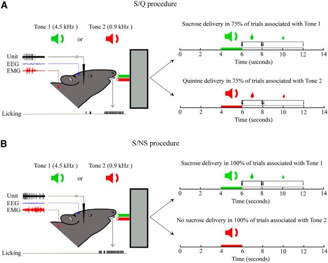
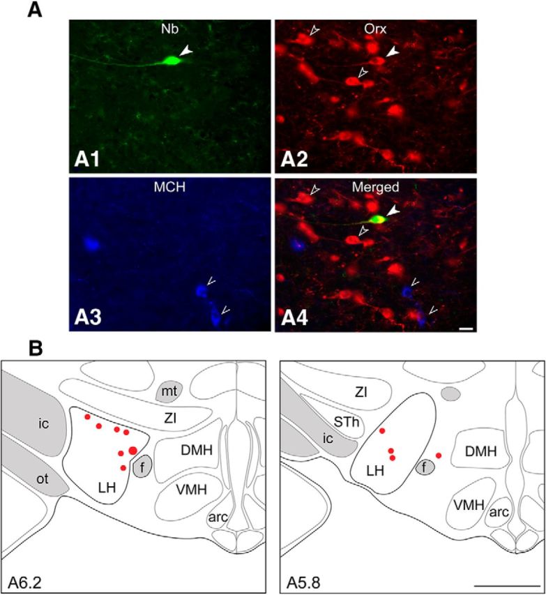
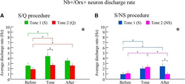
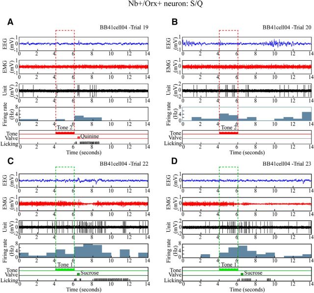
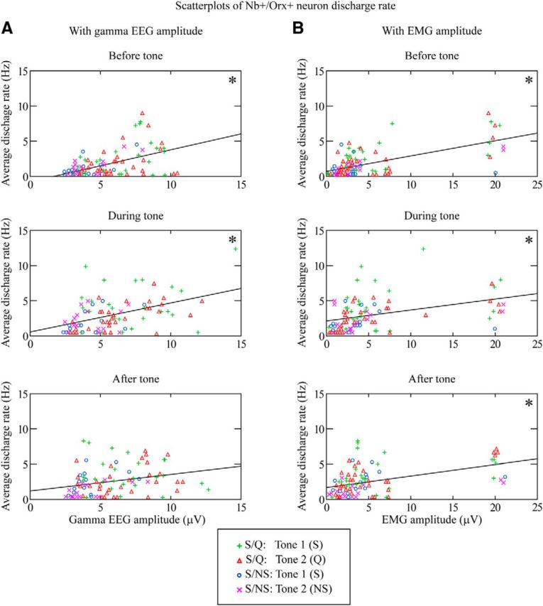
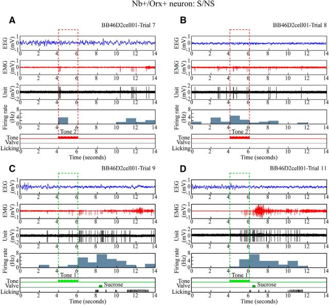
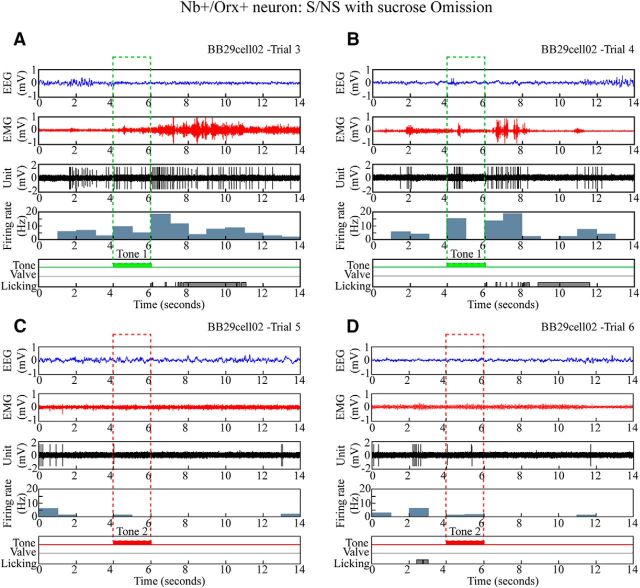
Orexin (Orx) neurons are known to be involved in the promotion and maintenance of waking because they discharge in association with cortical activation and muscle tone during waking and because, in their absence, waking with muscle tone cannot be maintained and narcolepsy with cataplexy ensues. Whether Orx neurons discharge during waking in association with particular conditions, notably with appetitive versus aversive stimuli or positive versus negative emotions, is debated and considered important in understanding their role in supporting particular waking behaviors. Here, we used the technique of juxtacellular recording and labeling in head-fixed rats to characterize the discharge of Orx neurons during the performance of an associative discrimination task with auditory cues for appetitive versus aversive outcomes. Of 57 active, recorded, and neurobiotin-labeled neurons in the lateral hypothalamus, 11 were immunohistochemically identified as Orx-positive (Orx(+)), whereas none were identified as melanin-concentrating hormone-positive. Orx(+) neurons discharged at significantly higher rates during the tone associated with sucrose than during the tone associated with quinine delivered upon licking. They also discharged at high rates after the tone associated with sucrose. Across periods and outcomes, their discharge was positively correlated with EEG gamma activity and EMG activity, which is indicative of cortical activation and behavioral arousal. These results suggest that Orx neurons discharge in a manner characteristic of reward neurons yet also characteristic of arousal neurons. Accordingly, the Orx neurons may respond to and participate in reward processes while modulating cortical activity and muscle tone to promote and maintain arousal along with learned adaptive behavioral responses.

Significance statement: Orexin neurons play a critical role in promoting and maintaining a waking state because, in their absence, narcolepsy with cataplexy ensues. Known to discharge during waking and not during sleep, they have also been proposed to be selectively active during appetitive behaviors. Here, we recorded and labeled neurons in rats to determine the discharge of immunohistochemically identified orexin neurons during performance of an associative discrimination task. Orexin neurons responded differentially to auditory cues associated with appetitive sucrose versus aversive quinine, indicating that they behave like reward neurons. However, correlated discharge with cortical and muscle activity indicates that they also behave like arousal neurons and can thus promote cortical activation with behavioral arousal and muscle tone during adaptive waking behaviors.

Keywords: EEG; EMG; MCH; arousal; reward; sleep.


Figures

Figure 1.
Figure 1.
Associative discrimination task. Rats were trained to respond to auditory stimuli of a high-frequency (4.5 kHz, Tone 1) or low-frequency (0.9 kHz, Tone 2) tone by licking to receive sucrose (S in A and B) after Tone 1 or not licking to avoid quinine (Q in A) or no sucrose (NS in B) after Tone 2. For data analysis, each behavioral trial was defined as a 14 s period that included a 4 s period before tone delivery, a 2 s period during tone, and an 8 s period after tone delivery. An interval of 10 ± 2 s occurred between these behavioral trials. To foster the rat's motivation and response, a bigger drop of sucrose was delivered if the animal responded within the initial 0–2 s window after the tone than if it responded within the subsequent 2–6 s window, after which no solution was delivered in response to licking. A, With the S/Q procedure, one group of animals was trained with partial reinforcement such that only 75% of sucrose-associated or quinine-associated trials were reinforced. B, With the S/NS procedure, another group of animals was trained to receive full reinforcement (100%) for sucrose-associated trials and no reinforcement (0%) for no sucrose-associated trials. The unit discharge was recorded together with EEG and EMG activity and licking. After recording and characterization over several trials, one neuron (per side per animal) was submitted to juxtacellular labeling with Nb for its immunohistochemical identification.
Figure 2.
Figure 2.
Identification and mapping of recorded, Nb+/Orx+ neurons. A, Fluorescence image of an Nb-labeled neuron (#BB41cell04, green, Cy2, filled arrowhead in A1) recorded from a rat submitted to the S/Q procedure, which was immunopositive for Orx (red, Cy3, filled arrowhead for Nb+/Orx+ and unfilled arrowheads for other Orx+ cells in A2 and A4) and negative for MCH (blue, Cy5, pointers in A3 and A4. Scale bar, 25 μm. B, Maps showing the location of Nb+/Orx+ recorded cells (as filled circles, with #BB41cell04 represented as the largest, at levels A ∼5.8 or 6.2 mm anterior to interaural zero) through the LH. Arc, Arcuate nucleus; DMH, dorsomedial hypothalamic nucleus; f, fornix; ic, internal capsule; mt, mammillo-thalamic tract; ot, optic tract; STh, subthalamic nucleus; VMH, ventromedial hypothalamic nucleus; ZI, zona incerta. Scale bar, 1 mm.
Figure 3.
Figure 3.
Average discharge rate of Nb+/Orx+ neurons with S/Q and S/NS procedures. The discharge of all Orx neurons varied significantly as a function of the period (before, during tone, and after), the tone (Tone 1 or 2), and the training (S/Q or S/NS). A, With the S/Q procedure (n = 8 cells, 57 trials), the discharge rate of Nb+/Orx+ neurons varied significantly across periods and tones. The average discharge rate during Tone 1 was significantly higher than that during Tone 2, reflecting discrimination of auditory cues associated with appetitive versus aversive valences. B, With the S/NS procedure (n = 3 cells, 29 trials), the discharge rate also varied significantly across periods and as a function of tone. The average discharge rate was significantly higher after Tone 1 (S) than after Tone 2 (NS), reflecting learned association with the receipt of appetitive sucrose. *p < 0.05 for period and or with tone in two-way mixed RM-ANOVAs for each procedure; *p < 0.05, according to post hoc comparisons; see text.
Figure 4.
Figure 4.
Discharge of Nb+/Orx+ neuron during S/Q discrimination task. Unit firing of representative Nb+/Orx+ neuron (#BB41cell04 illustrated in Fig. 2) shown along with EEG, EMG, and licking during four sequential Q and S trials. A, B, In quinine-rewarded trials, the neuron discharged at a low rate before the tone, minimally or moderately during Tone 2, and at a moderate rate after Tone 2 (Q), when the rat licked and received a quinine drop (valve) in one trial. The unit appeared to fire at higher rates in association with fast EEG activity. C, D, In sucrose-rewarded trials, the neuron discharged at a moderate rate before and higher rate during Tone 1 to reach its highest rate after Tone 1 (S), when the rat licked with short latency to receive a large drop of sucrose. Across all S and Q trials (n = 10) for this unit, the average discharge rate was positively correlated with gamma EEG amplitude (r = 0.401) and EMG amplitude (r = 0.693).
Figure 5.
Figure 5.
Correlations of Nb+/Orx+ neuron discharge with EEG gamma or EMG amplitude. Scatterplots are presented for the three periods (before tone, during tone, and after tone) and include data of individual trials (n = 86) from S/Q Tone 1 (S) (green plusses, 25), S/Q Tone 2 (Q) (red triangles, 32), S/NS Tone 1 (S) (blue circles, 14), and S/NS Tone 2 (NS) (magenta crosses, 15). A, As shown by the linear regression lines for all trials per period, the unit discharge rate was significantly positively correlated with gamma EEG amplitude during the before and tone periods. B, The unit discharge rate was also positively correlated with EMG amplitude during all three periods. *p < 0.05 according to Pearson's correlations with Dunn–Sidak-corrected probabilities; see text.
Figure 6.
Figure 6.
Discharge of Nb+/Orx+ neuron during S/NS discrimination task. Unit firing of representative Nb+/Orx+ neuron (#BB46D2cell01) shown along with EEG, EMG, and licking response during four sequential trials. A, B, In nonrewarded (no sucrose) Tone 2 trials, the unit discharged minimally before, moderately during Tone 2, and minimally after Tone 2 (NS), when the rat did not lick. C, D, In sucrose-rewarded Tone 1 trials, the unit discharged minimally before, moderately during Tone 1, and at a high rate after Tone 1 (S), when the rat licked to receive a small drop (in the later 2–6 s window in C) or large drop (within the initial 0–2 s window in D) of sucrose. The unit appeared to discharge at highest rates in association with fast EEG activity and EMG activity. Across all trials (n = 18), the average discharge rate was positively correlated with gamma EEG amplitude (r = 0.884) and EMG amplitude (r = 0.674).
Figure 7.
Figure 7.
Discharge of Nb+/Orx+ neuron during omission trials after training with S/NS procedure. The discharge of a representative Nb+/Orx+ (#BB29cell02) neuron is shown, along with the EEG, EMG, and licking response during four sequential trials. A, B, With expected sucrose-rewarded trials, the unit discharged at moderate rates before, increased rates during Tone 1, and high rates after Tone 1 (S), when the rat licked despite experimental omission of the sucrose. C, D, With expected nonrewarded trials of no sucrose, the unit discharged at moderate rates before and virtually not at all during Tone 2 and after Tone 2 (NS), when the rat did not lick. The unit appeared to discharge at its highest rates in association with fast EEG and EMG activity. Across trials (n = 8), the average discharge rate was positively correlated with gamma EEG amplitude (r = 0.659) and EMG amplitude (r = 0.467).

References

    1. Adamantidis AR, Zhang F, Aravanis AM, Deisseroth K, de Lecea L. Neural substrates of awakening probed with optogenetic control of hypocretin neurons. Nature. 2007;450:420–424. doi: 10.1038/nature06310. - DOI - PMC - PubMed
    1. Balcita-Pedicino JJ, Sesack SR. Orexin axons in the rat ventral tegmental area synapse infrequently onto dopamine and gamma-aminobutyric acid neurons. J Comp Neurol. 2007;503:668–684. doi: 10.1002/cne.21420. - DOI - PubMed
    1. Black SW, Morairty SR, Fisher SP, Chen TM, Warrier DR, Kilduff TS. Almorexant promotes sleep and exacerbates cataplexy in a murine model of narcolepsy. Sleep. 2013;36:325–336. - PMC - PubMed
    1. Blouin AM, Fried I, Wilson CL, Staba RJ, Behnke EJ, Lam HA, Maidment NT, Karlsson KÆ, Lapierre JL, Siegel JM. Human hypocretin and melanin-concentrating hormone levels are linked to emotion and social interaction. Nat Commun. 2013;4:1547. doi: 10.1038/ncomms2461. - DOI - PMC - PubMed
    1. Bonnavion P, Jackson AC, Carter ME, de Lecea L. Antagonistic interplay between hypocretin and leptin in the lateral hypothalamus regulates stress responses. Nat Commun. 2015;6:6266. doi: 10.1038/ncomms7266. - DOI - PMC - PubMed

Publication types