. 2016 Feb 3;36(5):1758-74.
doi: 10.1523/JNEUROSCI.3798-15.2016.

Pet-1 Switches Transcriptional Targets Postnatally to Regulate Maturation of Serotonin Neuron Excitability

Affiliations

Pet-1 Switches Transcriptional Targets Postnatally to Regulate Maturation of Serotonin Neuron Excitability

Steven C Wyler et al. J Neurosci. .

Abstract

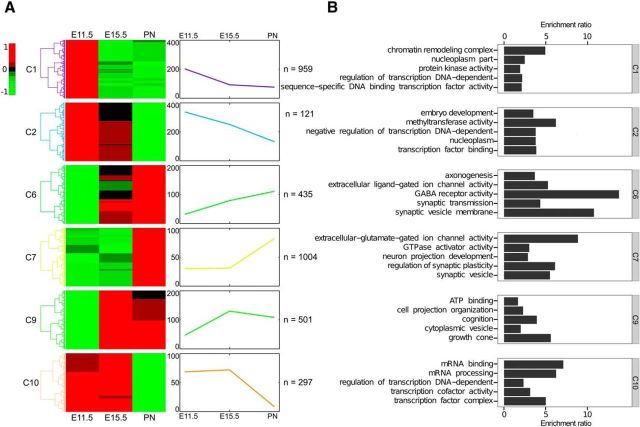
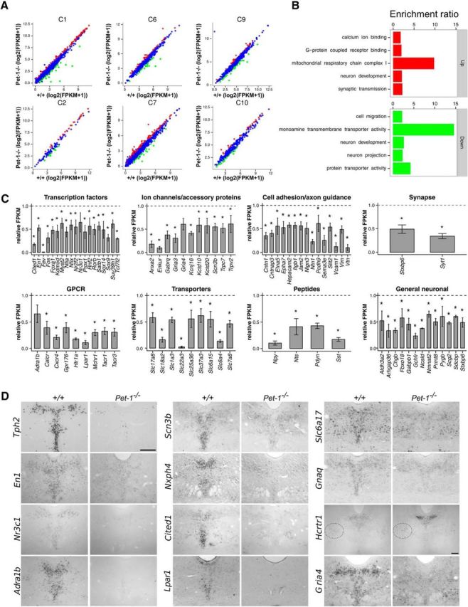
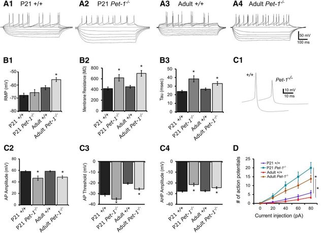
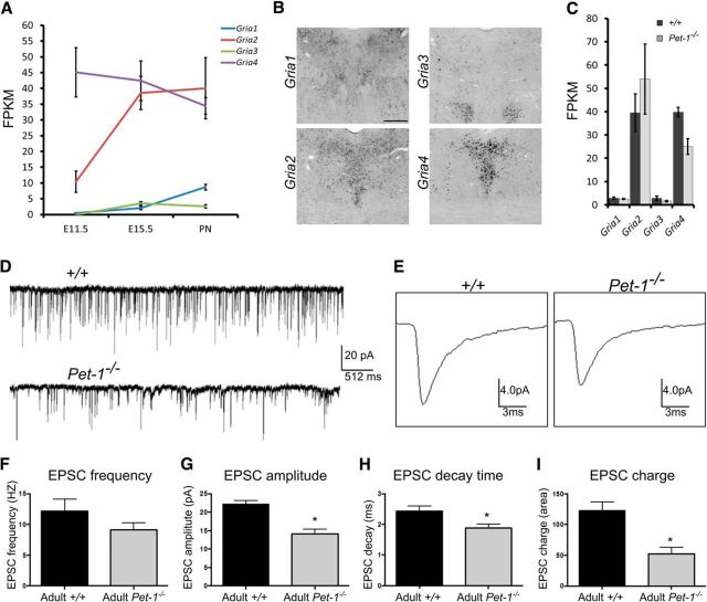
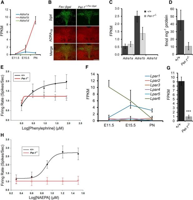
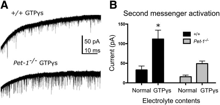
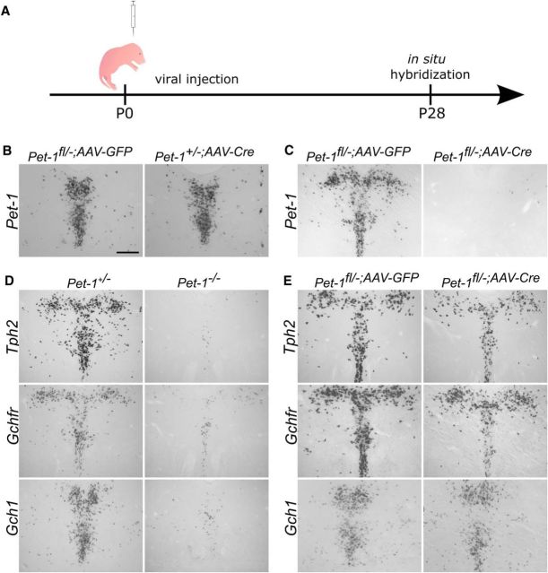
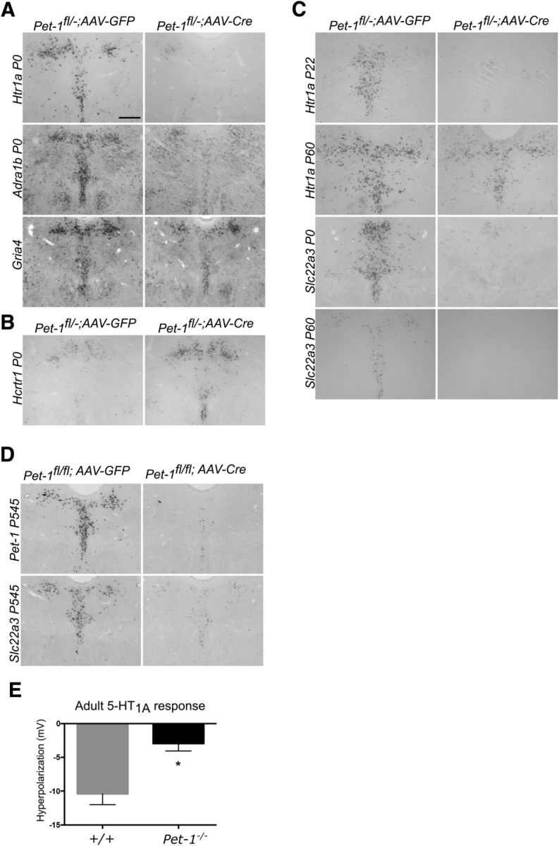
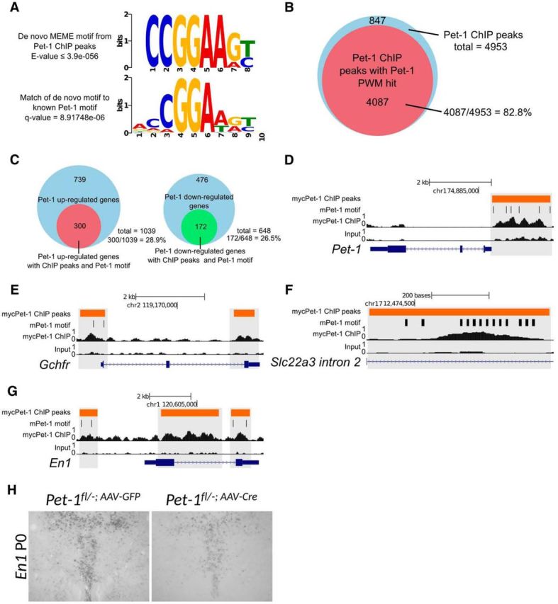
Newborn neurons enter an extended maturation stage, during which they acquire excitability characteristics crucial for development of presynaptic and postsynaptic connectivity. In contrast to earlier specification programs, little is known about the regulatory mechanisms that control neuronal maturation. The Pet-1 ETS (E26 transformation-specific) factor is continuously expressed in serotonin (5-HT) neurons and initially acts in postmitotic precursors to control acquisition of 5-HT transmitter identity. Using a combination of RNA sequencing, electrophysiology, and conditional targeting approaches, we determined gene expression patterns in maturing flow-sorted 5-HT neurons and the temporal requirements for Pet-1 in shaping these patterns for functional maturation of mouse 5-HT neurons. We report a profound disruption of postmitotic expression trajectories in Pet-1(-/-) neurons, which prevented postnatal maturation of 5-HT neuron passive and active intrinsic membrane properties, G-protein signaling, and synaptic responses to glutamatergic, lysophosphatidic, and adrenergic agonists. Unexpectedly, conditional targeting revealed a postnatal stage-specific switch in Pet-1 targets from 5-HT synthesis genes to transmitter receptor genes required for afferent modulation of 5-HT neuron excitability. Five-HT1a autoreceptor expression depended transiently on Pet-1, thus revealing an early postnatal sensitive period for control of 5-HT excitability genes. Chromatin immunoprecipitation followed by sequencing revealed that Pet-1 regulates 5-HT neuron maturation through direct gene activation and repression. Moreover, Pet-1 directly regulates the 5-HT neuron maturation factor Engrailed 1, which suggests Pet-1 orchestrates maturation through secondary postmitotic regulatory factors. The early postnatal switch in Pet-1 targets uncovers a distinct neonatal stage-specific function for Pet-1, during which it promotes maturation of 5-HT neuron excitability.

Significance statement: The regulatory mechanisms that control functional maturation of neurons are poorly understood. We show that in addition to inducing brain serotonin (5-HT) synthesis and reuptake, the Pet-1 ETS (E26 transformation-specific) factor subsequently globally coordinates postmitotic expression trajectories of genes necessary for maturation of 5-HT neuron excitability. Further, Pet-1 switches its transcriptional targets as 5-HT neurons mature from 5-HT synthesis genes to G-protein-coupled receptors, which are necessary for afferent synaptic modulation of 5-HT neuron excitability. Our findings uncover gene-specific switching of downstream targets as a previously unrecognized regulatory strategy through which continuously expressed transcription factors control acquisition of neuronal identity at different stages of development.

Keywords: Pet-1; adrenergic signaling; gene regulatory network; genomics; neuronal maturation; serotonin.


Figures

Figure 1.
Figure 1.
RNA-seq reveals temporal gene expression patterns in maturing 5-HT neurons. A, Total RNA from flow-sorted E11.5, E15.5, and P1–P3 (PN) YFP+ 5-HT neurons (n = 3 biological replicates/time point) was used for sequencing to determine expression patterns followed by hierarchical clustering of differentially expressed genes. Row-mean-normalized heat maps (left) and mean temporal expression levels (right) are shown for each cluster. Number of genes in each cluster is shown on the right of each trajectory. In total, 6126 genes were differentially expressed at ≥1.5-fold change and ≤5% FDR. B, GO enrichment analysis of gene expression clusters. GO terms enriched in clusters 6, 7, and 9 suggest increasing expression of genes related to neuronal maturation processes (hypergeometric test, p ≤ 0.01). GO terms enriched in clusters 1, 2, and 10 are associated with downregulation of earlier developmental programs involved in progenitor proliferation and specification (hypergeometric test, p ≤ 0.01).
Figure 2.
Figure 2.
RNA-seq shows that Pet-1 globally controls the 5-HT transcriptome through positive and negative regulation of gene-expression trajectories. A, Scatterplots showing altered expression of genes in Pet-1−/− versus +/+ 5-HT neurons in each expression cluster (Fig. 1). B, GO enrichment analysis of genes upregulated (top) and downregulated (bottom) by Pet-1. C, Relative changes in expression (FPKM) for various categories of Pet-1-controlled genes. *, Benjamini–Hochberg q value ≤0.05; n = 3. Error bars are SEM. D, ISH verification of genes regulated by Pet-1. Scale bar, 300 μm. Dotted oval shows nonserotonergic site of Hcrtr1 expression.
Figure 3.
Figure 3.
Pet-1−/− 5-HT neuron passive and active membrane properties are permanently immature. A, Whole-cell recordings of membrane voltage responses to hyperpolarizing and depolarizing current injection from P21 and adult +/+ and Pet-1−/− 5-HT neurons in the DRN. B, Passive membrane properties of Pet-1−/− 5-HT neurons are functionally immature. B1, Resting membrane potential (RMP; ANOVA F(3,97) = 9.177, p < 0.0001; n = 26, 11, 37, 27; Student–Newman–Keuls post hoc test: adult Pet-1−/− vs adult +/+ 5-HT neurons was significantly different). B2, Membrane resistance (ANOVA F(3,97) = 11.92, p < 0.0001; n = 26, 11, 37, 27; Student–Newman–Keuls t test: P21 and adult Pet-1−/− 5-HT neurons were significantly different from P21 and adult +/+ 5-HT neurons, respectively). B3, Membrane time constant (tau; ANOVA F(3,97) = 9.574, p < 0.0001; n = 26, 11, 37, 24; Student–Newman–Keuls t test: P21 and adult Pet-1−/− 5-HT neurons were significantly different from P21 and adult +/+ 5-HT neurons, respectively, p < 0.05. C, Persistent immaturity of AP characteristics in P21 and adult Pet-1−/− 5-HT neurons. C1, Representative raw data traces of an AP recorded from a P21 +/+ and a P21 Pet-1−/− 5-HT neuron. C2, AP amplitudes in P21 and adult +/+ and Pet-1−/− 5-HT neurons (ANOVA F(3,95) = 10.25, p < 0.0001; n = 26, 11, 35, 27; Student–Newman–Keuls t test confirmed that AP amplitude in P21 and adult Pet-1−/− 5-HT neurons was significantly smaller than P21 and adult +/+ 5-HT neurons, respectively. C3, Pet-1−/− 5-HT neuron AP firing threshold was significantly more hyperpolarized in P21 and remained more hyperpolarized in adult Pet-1−/− 5-HT neurons (F(3,95) = 37.92, p < 0.0001; n = 26, 11, 35, 27; Student–Newman–Keuls t test confirmed that P21 and adult Pet-1−/− 5-HT neurons more hyperpolarized than P21 and adult +/+ 5-HT neurons, respectively, p < 0.05). C4, Afterhyperpolarization amplitudes were smaller in P21 and adult Pet-1−/− 5-HT neurons (F(3,95) = 8.153, p < 0.0001; n = 26, 11, 35, 27; Student–Newman–Keuls test confirmed that P21 and adult Pet-1−/− 5-HT neurons were smaller than P21 and adult +/+ 5-HT neurons, respectively. D, Excitability of Pet-1−/− 5-HT neurons. Increased numbers of APs were elicited with depolarizing current pulses in P21 and adult Pet-1−/− 5-HT neurons compared with P21 and adult +/+ 5-HT neurons (two-way ANOVA significant interaction; ANOVA F(12,296) = 13.13, p < 0.0001; n = 26, 11, 17, 24).
Figure 4.
Figure 4.
Pet-1 promotes maturation of AMPA excitatory synaptic input to 5-HT neurons by regulating Gria4. A, RNA-seq analysis of AMPAR subunit gene (Gria1–4) expression trajectories (n = 3). B, ISH for Gria1–Gria4 in control mice. C, FPKMs for Gria1–4 in +/+ and Pet-1−/− sorted E15.5 5-HT neurons (n = 3). D–E, Raw traces of EPSC synaptic activity (D) and averaged (from 200 individual events) single EPSC current events (E) recorded under voltage-clamp conditions. F–I, EPSC input onto adult Pet-1−/− 5-HT neurons is characteristic of immature EPSCs at early postnatal stages. F, EPSC frequency was not different; however, the variances differed significantly (Pet-1−/−: 9.132 ± 1.125, N = 17; +/+: 13.75 ± 2.458, N = 22; F = 6.179, p < 0.0005; n = 22, 17). EPSC events in Pet-1−/− 5-HT neurons on average had smaller amplitudes (G; t test, t = 5.102, df = 37, p < 0.0001; n = 22, 17), shorter decay time (H; t test, t = 2.461, df = 37, p = 0.0186; n = 22, 17), and smaller charge (I; t test, t = 3.755, df = 37, p = 0.0006; n = 22, 17). Error bars are SEM.
Figure 5.
Figure 5.
Pet-1 controls maturation of GPCR synaptic input to 5-HT neurons. A, RNA-seq analysis of α1 adrenergic receptor gene-expression trajectories in flow-sorted +/+ 5-HT neurons. B, coimmunostaining of β-galactosidase (eFev::lacZ) marked 5-HT neurons (green) and ADRA1B (red). C, RNA-seq analysis of Adra1 receptor gene expression in +/+ and Pet-1−/− neurons at E15.5. D, [3H]-prazosin binding in +/+ versus Pet-1−/− midbrain (t(5) = 2.43, p = 0.07; n = 3. E, MEA recordings of α1-selective agonist phenylephrine (PE) responses. +/+, n = 19 cells/6 mice; Pet-1−/−, n = 24 cells/9 mice. F, RNA-seq analysis of Lpar1–6 expression trajectories in flow-sorted +/+ 5-HT neuron. G, FPKMs for Lpar1 in +/+ and Pet-1−/− sorted E15.5 5-HT neurons (n = 3). H, MEA recording of LPA1 selective agonist, (Z)-N-[2-(phosphonooxy)ethyl]-9-octadecenamide (NAEPA). +/+, n = 27 cells/4 mice; Pet-1−/−, n = 9 cells/3 mice. Scale bar, 300 μm. ***p < 0.001.
Figure 6.
Figure 6.
Immature G-protein signaling in Pet-1−/− 5-HT neurons. A, Representative image of GTPγS elicited responses in +/+ and Pet-1−/− 5-HT neurons. B, Quantification of GTPγS elicited response. ANOVA aCSF content F(1,31) = 13.59 p = 0.0009; genotype F(1,31) = 6.798, p = 0.0139; n = 4, 9, 6, 16. Student–Newman–Keuls t tests indicated that +/+ normal versus Pet-1−/− normal was not significantly different, +/+ normal versus +/+ GTPγS electrolyte was significant (*p < 0.05), and Pet-1−/− normal vs Pet-1−/− GTPγS was not significant.
Figure 7.
Figure 7.
5-HT synthesis genes lose sensitivity to Pet-1 as 5-HT neurons mature. A, Experimental scheme: Pet-1fl/− mice were injected with AAV-Cre or AAV-GFP to conditionally delete Pet-1 in the early postnatal period followed by ISH at P28. B, ISH for Pet-1fl/− and Pet-1+/− injected with AAV-GFP or AAV-Cre respectively. C, ISH for Pet-1 showing that AAV-Cre eliminates Pet-1 expression in the DRN. D, ISH reveals nearly complete elimination of expression of 5-HT synthesis genes Tph2, Gchfr, and Gch1 in the Pet-1−/− DRN. E, ISH in P0 AAV-injected mice reveals nearly total insensitivity of Tph2, Gchfr, and Gch1 expression to loss of Pet-1. Scale bar, 300 μm.
Figure 8.
Figure 8.
Early postnatal Pet-1 function is essential for control of multiple excitability genes. A, ISH of Htr1a, Adra1b, and, Gria4 expression in P0 injected mice. B, Neonatal targeting of Pet-1 results in increased Hcrtr1 expression in 5-HT neurons. C, Early postnatal transcriptional sensitive period for Htr1a control by Pet-1. Pet-1fl/− mice were injected with AAV-Cre or AAV-GFP at the indicated postnatal ages. ISH: Htr1a, P22 assayed at P43; Htr1a, P60 assayed at P180; Slc22a3, P0 assayed at P28; Slc22a3, P60 assayed at P90. D, ISH for Pet-1 and Slc22a3 of P545 injected mice assayed at P590. E, Permanent immaturity of 5-carboxamidotryptamine elicited responses in adult DRN slices. p = 0.0003, t(36) = 4.053. Pet-1−/−, n = 22; +/+, n = 16. Scale bar, 300 μm.
Figure 9.
Figure 9.
Pet-1 directly regulates the 5-HT neuron maturation factor Engrailed 1. A, De novo MEME motif analysis identifies the top significantly enriched motif in myc-Pet-1 peaks (top). TOMTOM Motif Comparison Tool identifies a highly significant match of the top enriched motif to Pet-1/FEV high-affinity binding site (bottom) defined in vitro (Wei et al., 2010). B, Fraction of mycPet-1 ChIP peaks with ≥1 match to the known Pet-1/FEV PWM motif. C, Fraction of Pet-1 upregulated (left) and downregulated (right) genes with mycPet-1 ChIP peaks within 5 kb from the TSS or TTS. D–G, Genome browser screen shots showing mycPet-1 enrichment over input control for Pet-1 (D), Gchfr (E), Slc22a3 (F), and En1 (G). Orange bars indicate area of significant peak enrichment. Black vertical lines indicate presence of Pet-1/FEV high-affinity motifs. H, P28 ISH for En1 of P0 Pet-1fl/− injected mice.

Similar articles

Cited by

References

    1. Allan DW, Thor S. Transcriptional selectors, masters, and combinatorial codes: regulatory principles of neural subtype specification. Wiley Interdiscip Rev Dev Biol. 2015;4:505–528. doi: 10.1002/wdev.191. - DOI - PMC - PubMed
    1. Baganz N, Horton R, Martin K, Holmes A, Daws LC. Repeated swim impairs serotonin clearance via a corticosterone-sensitive mechanism: organic cation transporter 3, the smoking gun. J Neurosci. 2010;30:15185–15195. doi: 10.1523/JNEUROSCI.2740-10.2010. - DOI - PMC - PubMed
    1. Bailey TL, Boden M, Buske FA, Frith M, Grant CE, Clementi L, Ren J, Li WW, Noble WS. MEME Suite: tools for motif discovery and searching. Nucleic Acids Res. 2009;37:W202–W208. doi: 10.1093/nar/gkp335. - DOI - PMC - PubMed
    1. Beck SG, Pan YZ, Akanwa AC, Kirby LG. Median and dorsal raphe neurons are not electrophysiologically identical. J Neurophysiol. 2004;91:994–1005. - PMC - PubMed
    1. Calizo LH, Akanwa A, Ma X, Pan YZ, Lemos JC, Craige C, Heemstra LA, Beck SG. Raphe serotonin neurons are not homogenous: electrophysiological, morphological and neurochemical evidence. Neuropharmacology. 2011;61:524–543. doi: 10.1016/j.neuropharm.2011.04.008. - DOI - PMC - PubMed

Publication types