Review
. 2016 Jan 27;8(1):65-76.
doi: 10.4240/wjgs.v8.i1.65.

Gallstone ileus, clinical presentation, diagnostic and treatment approach

Affiliations
Review

Gallstone ileus, clinical presentation, diagnostic and treatment approach

Carlos M Nuño-Guzmán et al. World J Gastrointest Surg. .

Abstract

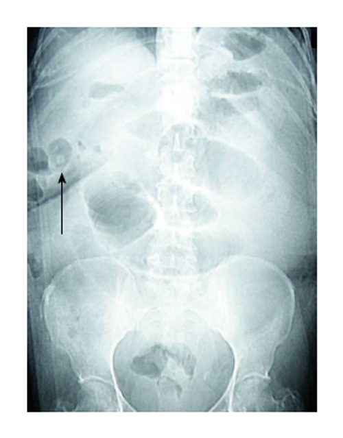
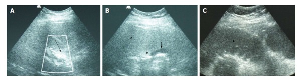
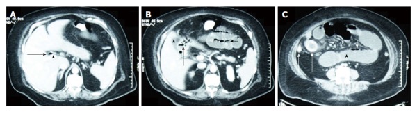
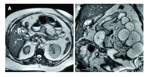
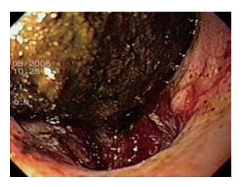
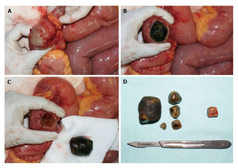
Gallstone ileus is a mechanical intestinal obstruction due to gallstone impaction within the gastrointestinal tract. Less than 1% of cases of intestinal obstruction are derived from this etiology. The symptoms and signs of gallstone ileus are mostly nonspecific. This entity has been observed with a higher frequency among the elderly, the majority of which have concomitant medical illness. Cardiovascular, pulmonary, and metabolic diseases should be considered as they may affect the prognosis. Surgical relief of gastrointestinal obstruction remains the mainstay of operative treatment. The current surgical procedures are: (1) simple enterolithotomy; (2) enterolithotomy, cholecystectomy and fistula closure (one-stage procedure); and (3) enterolithotomy with cholecystectomy performed later (two-stage procedure). Bowel resection is necessary in certain cases after enterolithotomy is performed. Large prospective laparoscopic and endoscopic trials are expected.

Keywords: Bouveret’s syndrome; Endoscopic treatment; Gallstone ileus; Intestinal obstruction; Laparoscopic surgery.


Figures

Figure 1
Figure 1
Plain abdominal radiograph showing dilated small bowel loops and a high density endoluminal image suggestive of a gallstone (arrow). No pneumobilia is visualized.
Figure 2
Figure 2
Ultrasound findings in a patient with gallstone ileus. A: Hyperechoic images without acoustic shadow in a non-dilated common bile duct, suggestive of air in the bile duct (arrow). Right portal vein was identified by Doppler US; B: US showing hyperechoic images without acoustic shadow in a collapsed gallbladder (arrow) and duodenum, suggestive of endoluminal air (short arrow). Liver parenchyma (arrowhead); C: Fluid-filled dilated proximal jejunum bowel loop (arrowhead). US: Ultrasound.
Figure 3
Figure 3
Contrast-enhanced computed tomography findings in a patient with gallstone ileus. A: Portal phase IV-contrast enhanced computed tomography section reveals air in the hepatic duct (arrow), anterior to a permeable right portal vein (arrowhead); B: Communication between a non-distended gallbladder (arrowhead) and the duodenum (arrow), where presence of air is observed. Fluid-filled dilated jejunum loops and intestinal pneumatosis are seen (short arrow); C: Endoluminal round-shaped calcium-density images (arrows), and dilated small bowel loops (arrowhead) with pneumatosis (short arrow).
Figure 4
Figure 4
Magnetic resonance cholangiopancreatography findings in a patient with gallstone ileus. A: On T2-MRI, a hyperintense image is identified in the gallbladder bed (arrow), with communication with the duodenal second portion (arrowhead), suggestive of a cholecystoduodenal fistula; B: MRI coronal reconstruction showed dilated small bowel loops with endoluminal air (black arrowheads) and a signal-void round-shaped image, suggestive of a gallstone (arrow). Gallbladder communication with duodenum is observed (white arrowhead). MRI: Magnetic resonance imaging.
Figure 5
Figure 5
Esophagogastroduodenoscopy in a patient with Bouveret’s syndrome revealed a gallstone in the duodenal bulb and the fistulous sinus. Courtesy of Gabriela Quintero-Tejeda, MD, Department of Gastrointestinal Endoscopy, Unidad Médica de Alta Especialidad, Hospital de Especialidades del Centro Médico Nacional de Occidente, Instituto Mexicano del Seguro Social.
Figure 6
Figure 6
Surgical findings in a patient with gallstone ileus. A: An impacted gallstone was found in distal jejunum. A smaller gallstone proximal to the impacted one is observed; B: Enterotomy over the site of the impacted gallstone; C: Intestinal wall compromise due to gallstone impaction can be observed; D: The offending gallstone, plus four of smaller dimensions found in proximal jejunum. An obstructing gallstone was found and extracted from the common bile duct in the same patient (gallstone on the right).

References

    1. Abou-Saif A, Al-Kawas FH. Complications of gallstone disease: Mirizzi syndrome, cholecystocholedochal fistula, and gallstone ileus. Am J Gastroenterol. 2002;97:249–254. - PubMed
    1. Martin F. Intestinal obstruction due to gall-stones: with report of three successful cases. Ann Surg. 1912;55:725–743. - PMC - PubMed
    1. Courvoisier LG. Casuistisch-statistische Beitrage zur Pathologic und Chirurgie der gallenwege. XII Leipzig, FCW Vogel: 1890.
    1. Bouveret L. Stenose du pylore adherent a la vesicule calculeuse. Rev Med (Paris) 1896;16:1–16.
    1. Kurtz RJ, Heimann TM, Beck AR, Kurtz AB. Patterns of treatment of gallstone ileus over a 45-year period. Am J Gastroenterol. 1985;80:95–98. - PubMed

LinkOut - more resources