

Efficiency of calcium phosphate composite nanoparticles in targeting Ehrlich carcinoma cells transplanted in mice
- PMID: 26843980
- PMCID: PMC4703481
- DOI: 10.1016/j.jare.2015.04.001
Efficiency of calcium phosphate composite nanoparticles in targeting Ehrlich carcinoma cells transplanted in mice
Abstract
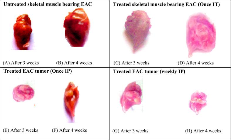
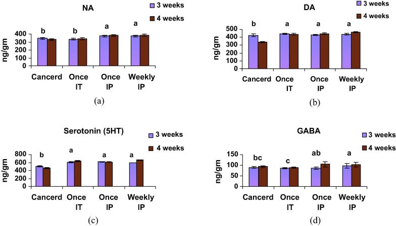
The present study aimed to investigate the mode of action of nano-CaPs in vivo as a therapy for solid tumor in mice. To achieve this goal, Ehrlich Ascites Carcinoma (EAC) was transplanted into 85 Swiss male albino mice. After nine days, the mice were divided into 9 groups. Groups 1 and 2 were allocated as the EAC control. Groups 3 and 4 were injected once intratumorally (IT) by nano-calcium phosphate (nano-CaP). Groups 5 and 6 received once intraperitoneal injection (IP) of nano-CaP. Groups 7, 8, and 9 received nano-CaP (IP) weekly. Blood samples and thigh skeletal muscle were collected after three weeks from groups 1, 3, 5, and 7 and after four weeks from groups 2, 4, 6, and 8. On the other hand, group 9 received nano-CaP (IP) for four weeks and lasted for three months to follow up the recurrence of tumor and to ensure the safety of muscle by histopathological analysis. Tumor growth was monitored twice a week throughout the experiment. DNA fragmentation of tumor cells was evaluated. In thigh tissue, noradrenaline, dopamine, serotonin (5HT), and gamma-aminobutyric acid (GABA) were measured. In serum, 8-Hydroxy-deoxyguanosine (8-OHDG), adenosine triphosphate (ATP), and vascular endothelial growth factor (VEGF) were analyzed. Histopathological and biochemical results showed a significant therapeutic effect of nano-CaP on implanted solid tumor and this effect was more pronounced in the animals treated IP for four weeks. This improvement was evident from the repair of fragmented DNA, the significant decrease of caspase-3, 8-OHDG, myosin, and VEGF, and the significant increase of neurotransmitters (NA, DA, 5HT, and GABA). Additionally, histopathological examination showed complete recovery of cancer cells in the thigh muscle after three months.
Keywords: 5HT, serotonin; 8-OHDG, 8-hydroxy-deoxyguanosine; ATP, adenosine triphosphate; Calcium phosphate (CaP) nanoparticles; DNA, deoxyribonucleic acid; EAC transplantation; EAC, Ehrlich Ascites Carcinoma; FAK, focal adhesion kinase; FTIR, Fourier transform infrared; GABA, gamma aminobutyric acid; IP, intraperitoneal; IT, intratumoral; MAPK, mitogen-activated protein kinase; Nano-CaP, nano calcium phosphate; Nanomedicine; Neurotransmitters; RIR, reference intensity ratio; SEM, scanning electron microscopy; Solid tumor; TEM, transmission electron microscope; VEGFR2, vascular endothelial growth factor receptor 2; XRD, X-ray diffraction.
Figures


Similar articles
-
Acanthus ilicifolius plant extract prevents DNA alterations in a transplantable Ehrlich ascites carcinoma-bearing murine model.World J Gastroenterol. 2007 Dec 28;13(48):6538-48. doi: 10.3748/wjg.v13.i48.6538. World J Gastroenterol. 2007. PMID: 18161924 Free PMC article.
-
Influence of halloysite nanotubes on the efficiency of Asparaginase against mice Ehrlich solid carcinoma.Saudi J Biol Sci. 2022 May;29(5):3626-3634. doi: 10.1016/j.sjbs.2022.02.058. Epub 2022 Mar 4. Saudi J Biol Sci. 2022. PMID: 35844382 Free PMC article.
-
Antitumor activity of a molecularly imprinted nanopreparation of 5-flurouracil against Ehrlich's carcinoma solid tumors grown in mice: Comparison to free 5-flurouracil.Chem Biol Interact. 2018 Nov 1;295:52-63. doi: 10.1016/j.cbi.2018.04.019. Epub 2018 Apr 17. Chem Biol Interact. 2018. PMID: 29678497
-
Ursolic acid inhibits tumor angiogenesis and induces apoptosis through mitochondrial-dependent pathway in Ehrlich ascites carcinoma tumor.Chem Biol Interact. 2013 Nov 25;206(2):153-65. doi: 10.1016/j.cbi.2013.09.004. Epub 2013 Sep 17. Chem Biol Interact. 2013. PMID: 24051192
-
Bone tissue engineering via nanostructured calcium phosphate biomaterials and stem cells.Bone Res. 2014 Sep 30;2:14017. doi: 10.1038/boneres.2014.17. eCollection 2014. Bone Res. 2014. PMID: 26273526 Free PMC article. Review.
Cited by
-
Therapeutic effect of Arthrocnemum machrostachyum methanolic extract on Ehrlich solid tumor in mice.BMC Complement Med Ther. 2020 May 24;20(1):153. doi: 10.1186/s12906-020-02947-y. BMC Complement Med Ther. 2020. PMID: 32448237 Free PMC article.
-
Fabrication of Calcium Sulfate Coated Selenium Nanoparticles and Corresponding In-Vitro Cytotoxicity Effects Against 4T1 Breast Cancer Cell Line.Avicenna J Med Biotechnol. 2021 Oct-Dec;13(4):201-206. Avicenna J Med Biotechnol. 2021. PMID: 34900146 Free PMC article.
-
Lichens-A Potential Source for Nanoparticles Fabrication: A Review on Nanoparticles Biosynthesis and Their Prospective Applications.J Fungi (Basel). 2021 Apr 12;7(4):291. doi: 10.3390/jof7040291. J Fungi (Basel). 2021. PMID: 33921411 Free PMC article. Review.
References
LinkOut - more resources
Full Text Sources
Other Literature Sources
Research Materials
Miscellaneous
