. 2016:2016:1752176.
doi: 10.1155/2016/1752176. Epub 2015 Dec 30.

Identification and Characterization of the V(D)J Recombination Activating Gene 1 in Long-Term Memory of Context Fear Conditioning

Affiliations

Identification and Characterization of the V(D)J Recombination Activating Gene 1 in Long-Term Memory of Context Fear Conditioning

Edgardo Castro-Pérez et al. Neural Plast. 2016.

Abstract

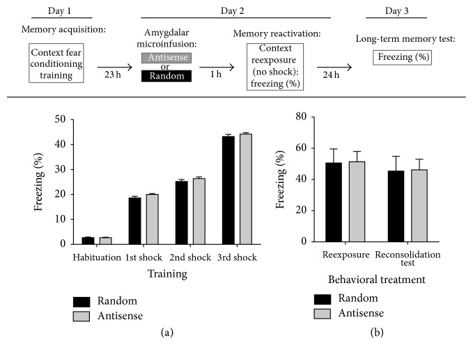
An increasing body of evidence suggests that mechanisms related to the introduction and repair of DNA double strand breaks (DSBs) may be associated with long-term memory (LTM) processes. Previous studies from our group suggested that factors known to function in DNA recombination/repair machineries, such as DNA ligases, polymerases, and DNA endonucleases, play a role in LTM. Here we report data using C57BL/6 mice showing that the V(D)J recombination-activating gene 1 (RAG1), which encodes a factor that introduces DSBs in immunoglobulin and T-cell receptor genes, is induced in the amygdala, but not in the hippocampus, after context fear conditioning. Amygdalar induction of RAG1 mRNA, measured by real-time PCR, was not observed in context-only or shock-only controls, suggesting that the context fear conditioning response is related to associative learning processes. Furthermore, double immunofluorescence studies demonstrated the neuronal localization of RAG1 protein in amygdalar sections prepared after perfusion and fixation. In functional studies, intra-amygdalar injections of RAG1 gapmer antisense oligonucleotides, given 1 h prior to conditioning, resulted in amygdalar knockdown of RAG1 mRNA and a significant impairment in LTM, tested 24 h after training. Overall, these findings suggest that the V(D)J recombination-activating gene 1, RAG1, may play a role in LTM consolidation.


Figures

Figure 1
Figure 1
Context fear conditioning induced upregulation of RAG1 mRNA in the amygdala. RAG1 mRNA levels were measured in the hippocampus and the amygdala on a time course at 15 min, 30 min, and 1 h after conditioning. (a) Normalized mRNA data showed no significant differences when examining hippocampal RAG1 mRNA Naïve (N) or the conditioned (C) groups sacrificed at 15, 30, or 60 min after training. (b) In contrast, context fear conditioning results in a significant, rapid, and transient induction in RAG1 mRNA levels in the amygdala (Naïve versus C15 min, P < 0.05; Naïve versus C30 min, # P < 0.05; Naïve versus C60 min, P > 0.05; C60 min versus C30 min, + P < 0.05; and C60 min versus C15 min, P > 0.05). (c) We sacrificed animals from the conditioned (C), CO, or SO groups 15 min after their respective associative or nonassociative training. Normalized expression confirmed the significant induction at 15 min of amygdalar RAG1 mRNA after context fear conditioning compared to Naïve, CO, and SO groups and showed no statistical difference between Naïve, CO, or SO controls (SO15 min versus C15 min, # P < 0.05; SO15 min versus Naive, P > 0.05; SO15 min versus CO15 min, P > 0.05; CO15 min versus C15 min, ++ P < 0.01; CO15 min versus Naive, P > 0.05; and Naïve versus C15 min ∗∗ P < 0.01).
Figure 2
Figure 2
RAG1 protein expression in amygdalar neuronal cells. Amygdalar coronal sections of context fear conditioning-trained mice, perfused 1 h after conditioning, were used for immunofluorescence and analyzed by confocal microscopy. Antibodies from immunofluorescence were validated by Western blot analysis. (a) Amygdalar area representative images of a double immunostaining using RAG1 antibody labeled with Alexa Fluor 488, green channel signal, and NeuN antibody labeled with Alexa Fluor 568, red channel signal. The left panel shows the NeuN positive neuronal nuclei, while the middle panel depicts RAG1 immunopositive cells. The right panel is the merge image showing colocalization of the NeuN neuronal nuclei marker and RAG1. Arrows point to some of the RAG1 immunopositive neurons. These immunofluorescent images revealed colocalization of RAG1 protein expressing cells with those expressing NeuN, suggesting the presence of RAG1 in neurons, although not all neurons expressed RAG1. (b) Tissue punches from amygdala (Amy) were obtained 1 h after context fear conditioning and analyzed in Western blot by comparative comigration with a standard molecular weight (MW) marker and protein extracts from bone marrow (BM) ((b)-1) and thymus (Thy) ((b)-2). Both sets of experiments consistently showed comigration between the tissues with a band corresponding to ~120 KD of RAG1 protein (green channel corresponding to RAG1 and red channel corresponding to beta-actin, ~42 KD); prestained molecular weight (MW) marker (ladder) was included in all the Western blots. ((b)-3) Additionally, tissue protein extracts from leg muscle (Mus) (negative control) were analyzed compared to amygdalar extracts with respect to RAG1 expression. As expected, RAG1 was not expressed in muscle compared to amygdala ((b)-3), bone marrow ((b)-1), and thymus ((b)-2). ((b)-4) RAG1 antibody preabsorption assays, either with muscle or with bone marrow extracts, showed that only bone marrow extracts, which express RAG1 as opposed to muscle, were able to block the ~120 KD band from amygdalar protein extracts in the Western blots, indicating that RAG1 antibody was preabsorbed (blocked) only by RAG1 protein expressing tissue (bone marrow).
Figure 3
Figure 3
Distribution of cannula placements and RAG1 antisense oligonucleotide diffusion within the amygdala. After behavioral treatments with RAG1 antisense or random oligonucleotides, animals were microinfused the next day with thionine to verify cannulae injectors' placement. Another set of animals was used to observe FITC-labeled RAG1 antisense diffusion. (a) Schematic representation of the amygdala at different rostrocaudal planes illustrating the position of cannulae injectors determined by thionine microinfusion. Injector tips for each cannula are represented by dark spots. (b) FITC-RAG1 antisense diffusion within the amygdalar complex; arrow indicates the injector's tip. (c) Schemes of coronal sections showing the diffusion of FITC-RAG1 antisense diffusion into the amygdala of animals decapitated 3 h after fluorescent oligonucleotide infusion. FITC-RAG1 antisense diffusion is represented by green shading from anterior to posterior areas of the amygdalar complex. The numbers in (a) and (c) indicate the distance from bregma in millimeters. A total of 4 mice were used in these studies. (d) Photomicrograph at higher magnification of FITC-RAG1 antisense diffusion showed clearly incorporation into the cells (depicted by the arrows) within amygdalar regions.
Figure 4
Figure 4
RAG1 antisense amygdalar treatment impaired consolidation of context fear conditioning. Top panel: diagram depicting the experimental design of these experiments for pretraining or posttraining amygdalar antisense or random oligonucleotide microinfusion experiments. In the pretraining microinfusion experiments, mice received RAG1 antisense or random bilateral oligonucleotide microinfusions directed at the amygdala 1 h before conditioning followed by either LTM testing or molecular evaluation. LTM was tested 24 h after conditioning. For molecular evaluation of antisense treatment effectiveness, another group of mice was sacrificed 30 min after conditioning and amygdalar RNA was used for real-time PCR. In the posttraining microinfusion experiments, mice were conditioned, returned to their home cages, and received microinfusions of antisense or random oligonucleotides 5 h after training and returned to their home cages until next day. Nineteen (19) hours later (24 h after conditioning), mice were reexposed to the conditioning chamber without any shocks in order to test LTM. (a) Mice receiving either RAG1 antisense or random oligonucleotide treatment displayed no significant differences during memory acquisition measured as the progressive enhancement of freezing behavior (Two-Way ANOVA, Treatment Factor: F(1,0.8457) = 0.01015, P > 0.9; Training Factor F(3,7863) = 94.37, ∗∗∗ P < 0.0001; Interaction: F(3,7.457) = 0.08950, P > 0.9). Bonferroni posttesting analysis did not identify significant differences between the groups during the habituation or the 1st, 2nd, or 3rd trials of training (P > 0.05, each comparison), indicating that both groups were similarly capable of learning the task. (b) LTM was tested 24 h after conditioning. The bar graph shows that, unlike the results obtained for acquisition, mice treated with RAG1 antisense gapmer oligonucleotides displayed significantly less percent freezing to the conditioning context than random oligonucleotide controls during the LTM test (Student's t-test; t (25) = 2.602; P < 0.05). (c) The molecular effectiveness of our knockdown by gapmer antisense oligonucleotide of RAG1 in the amygdala was determined by quantitative real-time PCR. Mice were infused 1 h before context fear conditioning with bilateral RAG1 antisense or random oligonucleotides and decapitated 30 min after conditioning. RAG1 mRNA normalized against gapdh mRNA showed that treatment with RAG1 antisense gapmer oligonucleotides effectively knocked down the levels of RAG1 amygdalar mRNA compared to the random controls (Student's t-test; t (16) = 3.947; ∗∗ P < 0.005). No significant differences in the levels of gapdh were observed between treatments (data not shown). (d) We used 5 h posttraining amygdalar microinfusions of RAG1 antisense oligonucleotides or random controls with a different set of animals without any pretraining infusion and LTM was tested 24 h after training. Unlike in the pretraining microinfusion experiments, both the antisense and random posttraining-infused mice displayed similar levels of conditioned freezing during the LTM test (Student's t-test; t (12) = 2.835; P > 0.7).
Figure 5
Figure 5
RAG1 antisense amygdalar treatment does not interfere with reconsolidation of context fear conditioning. To test the effects of RAG1 gapmer antisense treatment on memory reconsolidation of context fear conditioning, another set of animals was bilaterally implanted with cannulas to target the amygdala. Top panel: diagram depicting the experimental design. On day 1, mice were trained in context fear conditioning and immediately returned to their home cages. Antisense or random oligonucleotides were microinfused into the amygdala 1 h before memory reactivation on day 2. The effect of antisense or random oligonucleotide treatment on LTM reconsolidation was assessed on day 3, 48 h after conditioning. (a) On day 1, mice were microinfused with saline 1 h before training and returned to their home cages immediately after conditioning. Two-Way RM ANOVA and Bonferroni posttesting demonstrated that the infusions did not impair the animals' response in developing and expressing fear during the conditioning experience (Treatment Assignment Factor: F(1,8.194) = 3.979, P > 0.05; Training Factor F(3,3134) = 1725, ∗∗∗ P < 0.0001; and Interaction: F(3,1.069) = 0.5881, P > 0.6). (b) On day 2, animals were microinfused with either random or antisense gapmer oligonucleotides 1 h prior to a 90 s reexposure period to the conditioning chamber in order to induce memory retrieval and returned to their home cages. For the reconsolidation test, on day 3 (48 h after training), mice were reexposed to the conditioning chamber (CS) for 2 min to measure freezing responses. No significant differences between the freezing responses of antisense or random gapmer oligonucleotide treated animals on day 2 (b) or on day 3 (b) were observed (Two-Way ANOVA: Treatment Factor: F(1,3.068) = 0.009017, P > 0.9; Training Factor F(1,146.5) = 0.4307, P > 0.5; Interaction: F(1,0.001515) = 0.00004453, P > 0.9). Bonferroni posttesting identified no difference between treatments in the reexposure and reconsolidation tests, respectively (P > 0.05, each comparison).

Similar articles

Cited by

References

    1. Baranes D., Lederfein D., Huang V.-Y., Chen M., Bailey C. H., Kandel E. R. Tissue plasminogen activator contributes to the late phase of LTP and to synaptic growth in the hippocampal mossy fiber pathway. Neuron. 1998;21(4):813–825. doi: 10.1016/s0896-6273(00)80597-8. - DOI - PubMed
    1. Maletic-Savatic M., Malinow R., Svoboda K. Rapid dendritic morphogenesis in CA1 hippocampal dendrites induced by synaptic activity. Science. 1999;283(5409):1923–1927. doi: 10.1126/science.283.5409.1923. - DOI - PubMed
    1. Toni N., Buchs P.-A., Nikonenko I., Bron C. R., Muller D. LTP promotes formation of multiple spine synapses between a single axon terminal and a dendrite. Nature. 1999;402(6760):421–425. doi: 10.1038/46574. - DOI - PubMed
    1. Goshen I., Brodsky M., Prakash R., et al. Dynamics of retrieval strategies for remote memories. Cell. 2011;147(3):678–689. doi: 10.1016/j.cell.2011.09.033. - DOI - PubMed
    1. Davis H. P., Squire L. R. Protein synthesis and memory: a review. Psychological Bulletin. 1984;96(3):518–559. doi: 10.1037/0033-2909.96.3.518. - DOI - PubMed

Publication types

LinkOut - more resources