. 2016 Mar:33:13-24.
doi: 10.1016/j.actbio.2016.01.043. Epub 2016 Feb 1.

Extracellular matrix mediators of metastatic cell colonization characterized using scaffold mimics of the pre-metastatic niche

Affiliations

Extracellular matrix mediators of metastatic cell colonization characterized using scaffold mimics of the pre-metastatic niche

Brian A Aguado et al. Acta Biomater. 2016 Mar.

Abstract

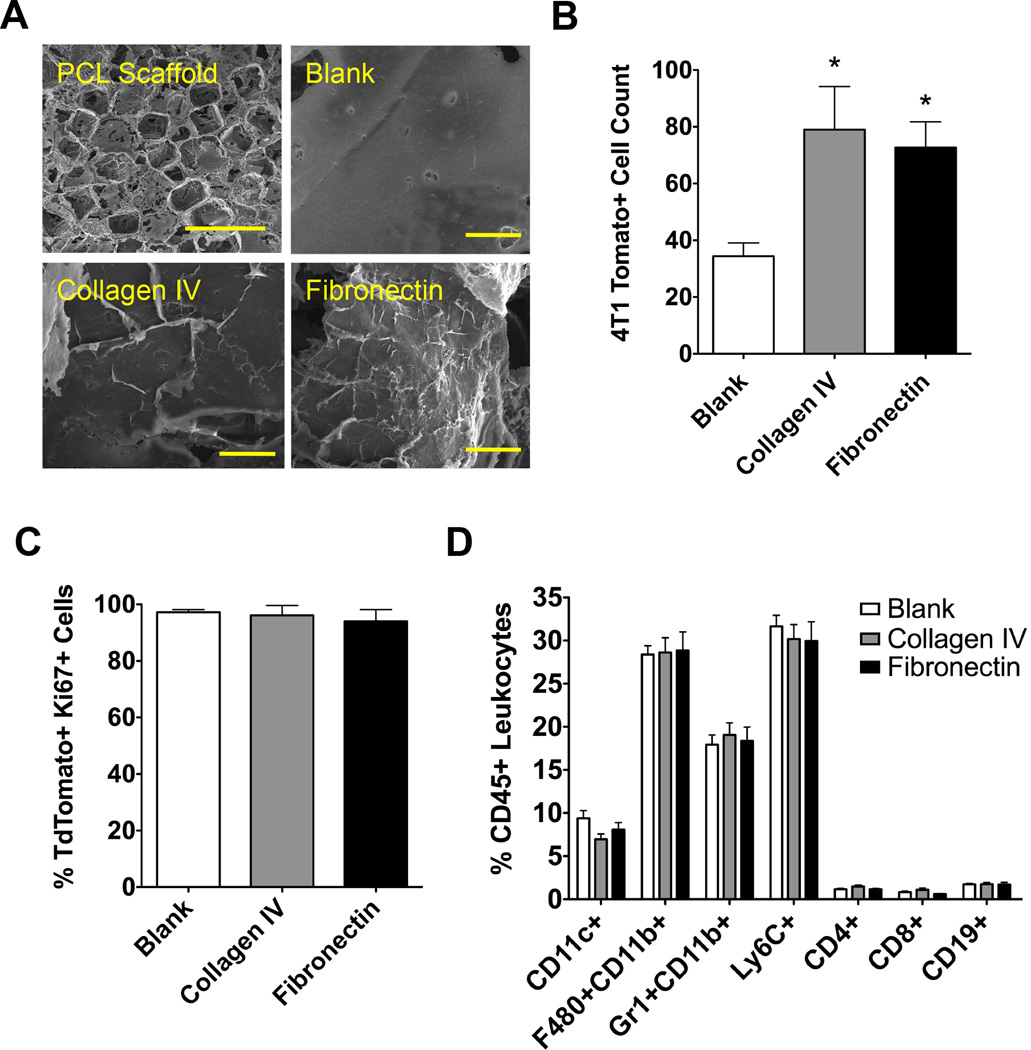
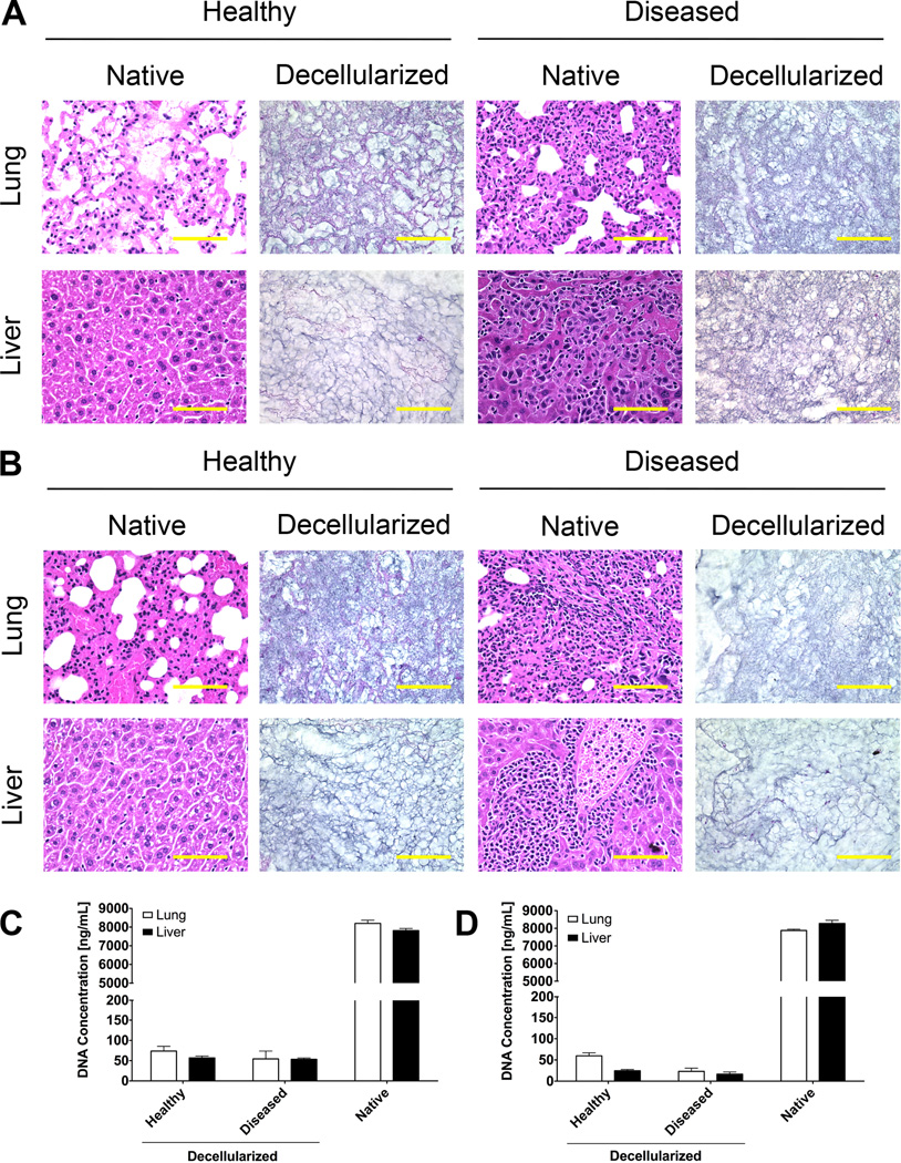
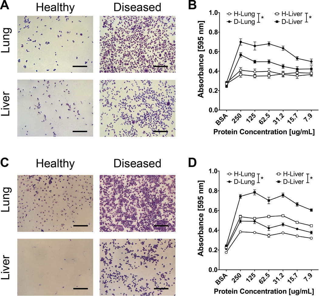
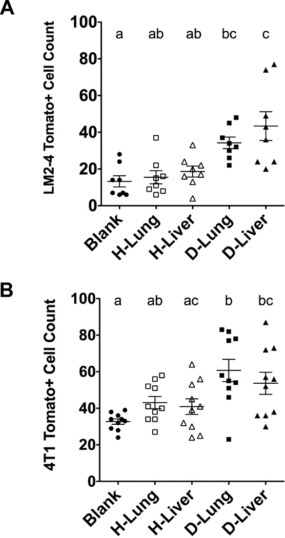
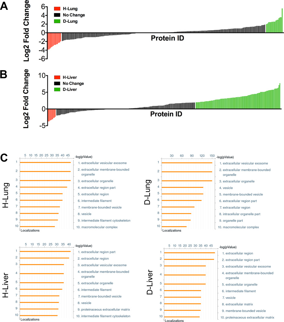
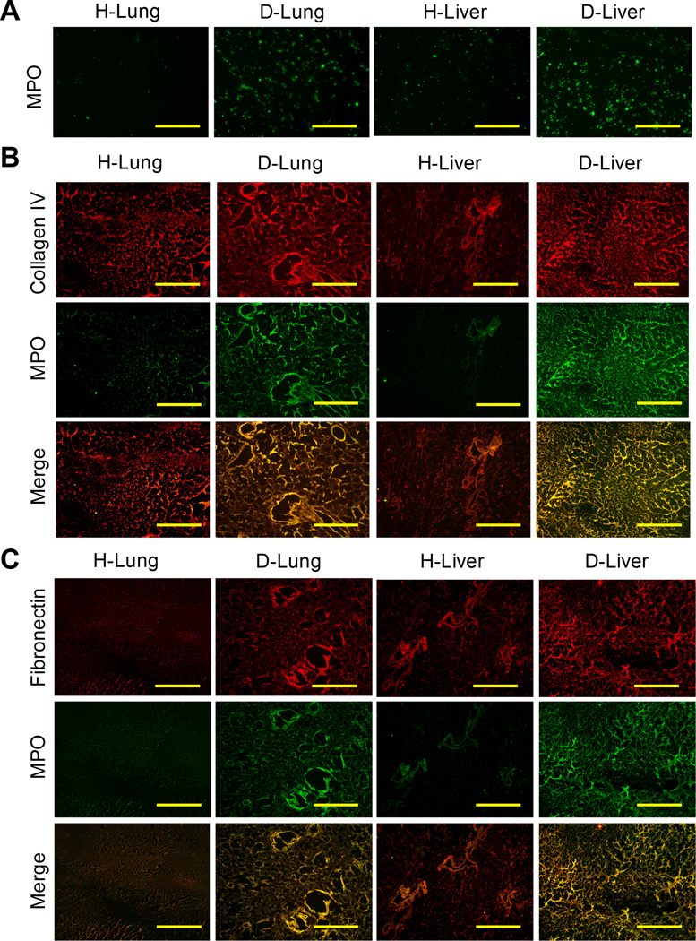
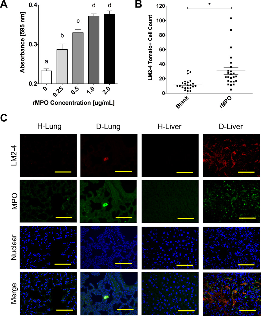
Metastatic tumor cells colonize the pre-metastatic niche, which is a complex microenvironment consisting partially of extracellular matrix (ECM) proteins. We sought to identify and validate novel contributors to tumor cell colonization using ECM-coated poly(ε-caprolactone) (PCL) scaffolds as mimics of the pre-metastatic niche. Utilizing orthotopic breast cancer mouse models, fibronectin and collagen IV-coated scaffolds implanted in the subcutaneous space captured colonizing tumor cells, showing a greater than 2-fold increase in tumor cell accumulation at the implant site compared to uncoated scaffolds. As a strategy to identify additional ECM colonization contributors, decellularized matrix (DCM) from lungs and livers containing metastatic tumors were characterized. In vitro, metastatic cell adhesion was increased on DCM coatings from diseased organs relative to healthy DCM. Furthermore, in vivo implantations of diseased DCM-coated scaffolds had increased tumor cell colonization relative to healthy DCM coatings. Mass-spectrometry proteomics was performed on healthy and diseased DCM to identify candidates associated with colonization. Myeloperoxidase was identified as abundantly present in diseased organs and validated as a contributor to colonization using myeloperoxidase-coated scaffold implants. This work identified novel ECM proteins associated with colonization using decellularization and proteomics techniques and validated candidates using a scaffold to mimic the pre-metastatic niche.

Statement of significance: The pre-metastatic niche consists partially of ECM proteins that promote metastatic cell colonization to a target organ. We present a biomaterials-based approach to mimic this niche and identify ECM mediators of colonization. Using murine breast cancer models, we implanted microporous PCL scaffolds to recruit colonizing tumor cells in vivo. As a strategy to modulate colonization, we coated scaffolds with various ECM proteins, including decellularized lung and liver matrix from tumor-bearing mice. After characterizing the organ matrices using proteomics, myeloperoxidase was identified as an ECM protein contributing to colonization and validated using our scaffold. Our scaffold provides a platform to identify novel contributors to colonization and allows for the capture of colonizing tumor cells for a variety of downstream clinical applications.

Keywords: Colonization; Extracellular matrix; Metastasis; Organ decellularization; Pre-metastatic niche.


Figures

Figure 1
Figure 1. ECM-coated scaffolds for metastatic tumor cell colonization
(A) SEM images of a porous PCL scaffold (top left scale bar = 1 mm) with zoomed in details of an uncoated blank scaffold and coated collagen IV and fibronectin scaffolds (scale bars = 50 µm). (B) Flow cytometric analysis of 4T1 tumor cell recruitment to coated PCL scaffolds implanted subcutaneously. Asterisk indicates statistical significance compared to blank control, P < 0.05 (n = 10). (C) Percent of Ki67-positive tumor cells present at implanted scaffolds analyzed using flow cytometry (n = 10). (D) Flow cytometry quantification of immune cell populations at ECM-coated scaffolds (n = 10).
Figure 2
Figure 2. Decellularization of lungs and livers of healthy and diseased mice
H&E sections of healthy and diseased lungs and livers from (A) NSG mice inoculated with LM2-4 cells and (B) BalbC mice inoculated with 4T1 cells (scale bar = 100 µm). DNA concentrations of decellularized matrix and native tissue digests of (C) NSG and (D) BalbC lungs and livers (n = 4).
Figure 3
Figure 3. In vitro adhesion of LM2-4 and 4T1 cells on DCM coatings
(A) Representative images of LM2-4 cells adhered to tissue culture plastic coated with lung and liver DCM from healthy and diseased NSG mice (scale bars = 300 µm, protein concentration of coating solution = 250 µg/mL). (B) Crystal violet absorbance values of LM2-4 cells adhered on DCM matrix coatings (n = 12), asterisk indicates P < 0.05 comparison at each protein concentration value (excluding BSA control). (C) Representative images of 4T1 cells adhered to tissue culture plastic coated with lung and liver DCM from healthy and diseased BalbC mice (scale bars = 300 µm, protein concentration of coating solution = 250 µg). (D) Crystal violet absorbance values of 4T1 cells adhered on DCM matrix coatings (n = 12), asterisk indicates P < 0.05 comparison at each protein concentration value (excluding BSA control).
Figure 4
Figure 4. In vivo tumor cell colonization to PCL scaffolds coated with various DCM solutions implanted subcutaneously
(A) LM2-4 tumor cell colonization in NSG mice (n = 8) and (B) 4T1 tumor cell colonization in BalbC mice (n = 10) show increased tumor cell recruitment on diseased lung and liver DCM coatings compared to blank scaffolds. Groups with different letters indicate statistical significance (P < 0.05).
Figure 5
Figure 5. Proteomic characterization of DCM coatings
Graph showing log2 fold changes of healthy and diseased (A) lungs and (B) livers peptide spectral matches, where each tick on the x-axis represents an identified protein in the matrix. Log2 fold change values shaded in red indicate proteins 2-fold more abundant in healthy DCM, and log2 fold change values shaded in green indicate proteins 2-fold more abundant in diseased DCM. (C) Gene ontology analysis of lung and liver DCM coatings using MetaCore software show protein localizations.
Figure 6
Figure 6. Presence of myeloperoxidase in decellularized lung and liver tissues
(A) Confirmation of increased abundance of myeloperoxidase in diseased lung and liver DCM compared to healthy DCM using immunofluorescence stains of coatings on tissue culture plastic (scale bars = 100 µm). Co-immunofluorescence stains for (B) collagen IV or (C) fibronectin with myeloperoxidase, showing increased abundance of myeloperoxidase in diseased tissues (scale bars = 100 µm).
Figure 7
Figure 7. Validation of myeloperoxidase as a contributor to metastatic cell colonization
(A) Adhesion assay absorbance values showing concentration dependent adhesion of LM2-4 cells on tissue culture plastic treated with myeloperoxidase solutions at indicated protein concentrations (n = 4). Groups with different letters indicate statistical significance (P < 0.05). (B) In vivo colonization of LM2-4 cells on MPO-coated PCL scaffolds 7 days post subcutaneous implantation (n = 24). Asterisk indicates significance at P < 0.002. (D) Immunofluorescence stains showing MPO localization near tumor cell clusters in D-Lung and D-Liver sections compared to H-Lung and H-Liver controls (scale bars = 100 µm).

References

    1. Fidler IJ. The pathogenesis of cancer metastasis: the “seed and soil” hypothesis revisited. Nat. Rev. Cancer. 2003;3:1–6. - PubMed
    1. Nguyen DX, Bos PD, Massagué J. Metastasis: from dissemination to organ-specific colonization. Nat. Rev. Cancer. 2009;9:274–284. - PubMed
    1. Psaila B, Lyden D. The metastatic niche: adapting the foreign soil. Nat. Rev. Cancer. 2009;9:285–293. - PMC - PubMed
    1. Kaplan RN, Riba RD, Zacharoulis S, Bramley AH, Vincent L, Costa C, et al. VEGFR1-positive haematopoietic bone marrow progenitors initiate the pre-metastatic niche. Nature. 2005;438:820–827. - PMC - PubMed
    1. Wculek SK, Malanchi I. Neutrophils support lung colonization of metastasis-initiating breast cancer cells. Nature. 2015;528:413–417. - PMC - PubMed

Publication types

MeSH terms