. 2016 Feb 3;89(3):480-93.
doi: 10.1016/j.neuron.2015.12.026.

Dscam Proteins Direct Dendritic Targeting through Adhesion

Affiliations

Dscam Proteins Direct Dendritic Targeting through Adhesion

Wael Tadros et al. Neuron. .

Abstract

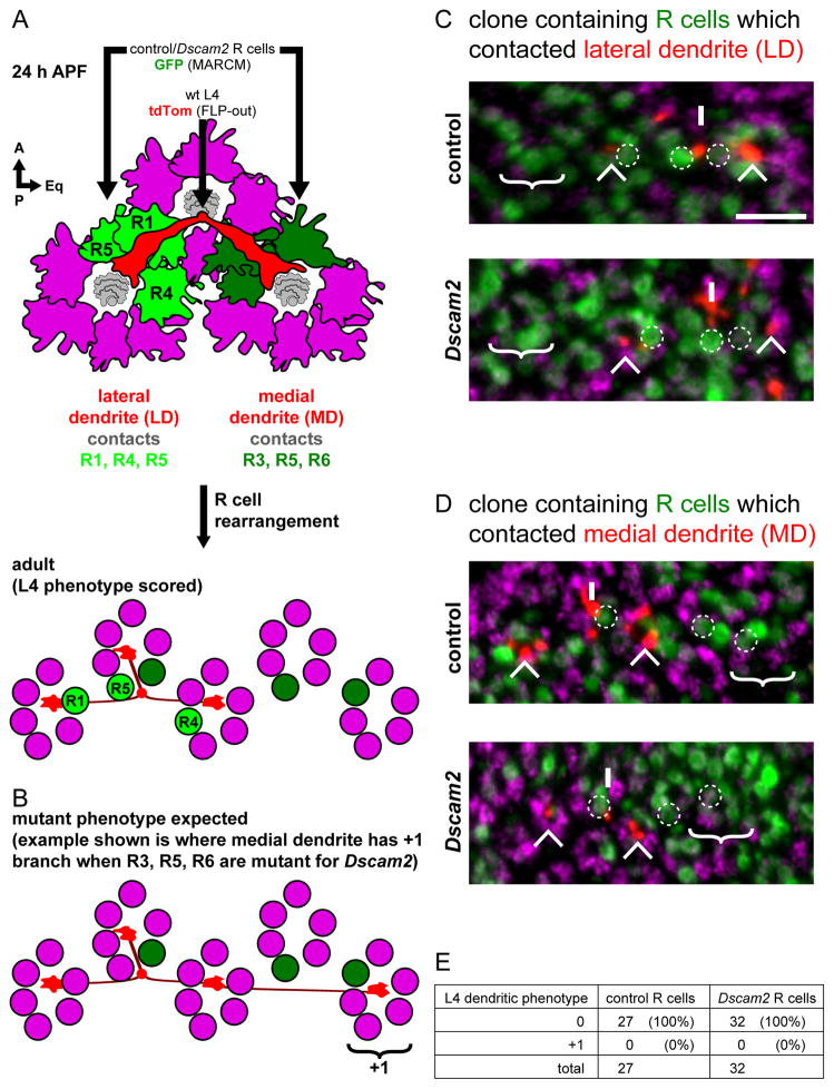
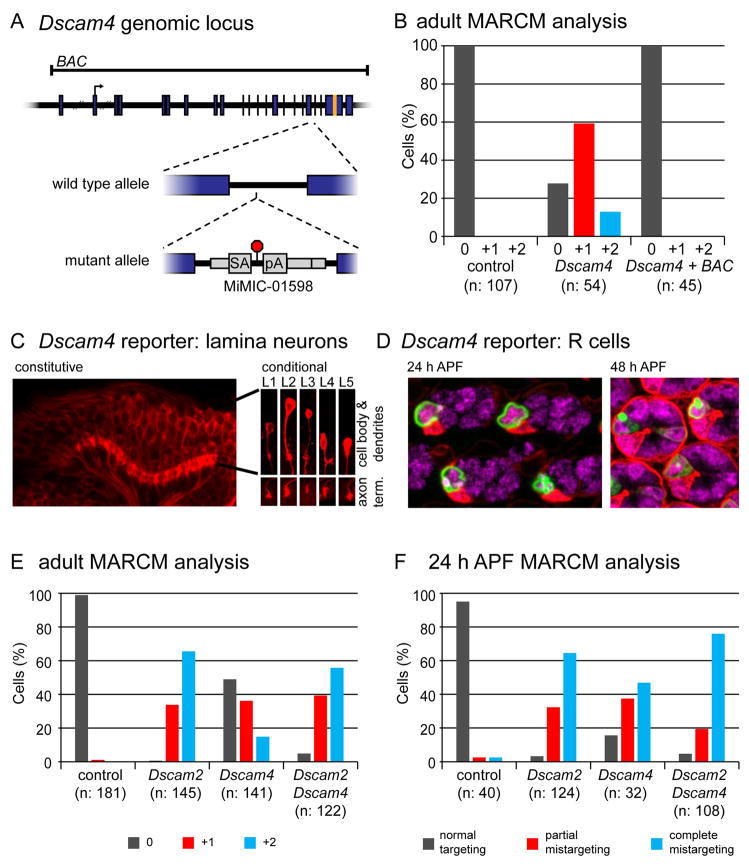
Cell recognition molecules are key regulators of neural circuit assembly. The Dscam family of recognition molecules in Drosophila has been shown to regulate interactions between neurons through homophilic repulsion. This is exemplified by Dscam1 and Dscam2, which together repel dendrites of lamina neurons, L1 and L2, in the visual system. By contrast, here we show that Dscam2 directs dendritic targeting of another lamina neuron, L4, through homophilic adhesion. Through live imaging and genetic mosaics to dissect interactions between specific cells, we show that Dscam2 is required in L4 and its target cells for correct dendritic targeting. In a genetic screen, we identified Dscam4 as another regulator of L4 targeting which acts with Dscam2 in the same pathway to regulate this process. This ensures tiling of the lamina neuropil through heterotypic interactions. Thus, different combinations of Dscam proteins act through distinct mechanisms in closely related neurons to pattern neural circuits.


Figures

Figure 1
Figure 1. Dscam2 is required for dendritic patterning of L4 neurons
(A) Schematic of the retina (R) and lamina (L) of the Drosophila visual system showing the location and arrangement of L4 neurons (green). A single L4, highlighted by the dotted box is detailed in (B). (B) Schematic of L4 neuron (green) innervating two posterior (away) cartridges containing R cells (red) and lamina neurons (grey). Home cartridge (see C) omitted for clarity. (C) Cross-sectional view of B (including home and away cartridges). Positions of R1-R6 and lamina neurons (L1-L5) indicated. Anterior (A), posterior (P), dorsal (D), ventral (V) and equatorial (Eq) orientations as indicated. The equator is the midline of the D-V axis. (D) L4 dendrites tile the lamina neuropil. (E) Schematic of Dscam2 genomic locus and mutant alleles. Location of translational start site (arrow), exons (blue boxes) and sequences encoding the transmembrane domain (TM) are indicated. Variable exons 10A and 10B in Dscam2 are demarcated in orange and pink, respectively (see also Figure S2A) Dscam2 null allele is a replacement of exon 1 with exogenous DNA bearing the white (w) gene (purple box; Millard et al., 2007). Isoform specific stop mutants, Dscam2Astop, Dscam2Bstop and Dscam2A+Bstop contain stop codons in exons 10A, 10B or both, respectively. (F) MARCM analysis of Dscam2 in adult L4 neurons. 3D renderings (top 3 panels) taken from serial confocal slices (bottom 3 panels) of wild-type and Dscam2 mutant L4 neurons. L4 neurons were labeled with GFP (green) and cartridges with anti-Highwire (Hiw; red). Dotted line indicates position of corresponding confocal slice. Axons (Ax; arrowheads) and additional dendritic branches (*) are indicated. Scale bar, 5 μm. (G) Quantification of dendritic phenotypes seen in MARCM-generated L4 neurons with Dscam2 null mutation. (H) Quantification of dendritic phenotypes seen in MARCM-generated L4 neurons with Dscam2 isoform-specific alleles.
Figure 2
Figure 2. L4 dendrites project to their target fascicles at an early stage in their development
(A,B) Dendrites from neighboring L4 neurons contact each other as they converge on a common target fascicle at 24 h APF. (A) Schematic representation of two adjacent L4 neurons (green and blue) imaged in (B). A lattice of R cell growth cones (red) surrounds each lamina fascicle (purple). Glia (grey) lie immediately beneath L4 dendrites. Dotted line indicates the cross-section at which the side view is taken. (B) Multicolor Flip Out (MCFO; Nern et al., 2015) was used to simultaneously label two adjacent L4s. Confocal sections (top panel; MIP: maximum intensity projection) were used to generate 3D renderings of L4 neurons and R cell growth cones displayed from different perspectives (bottom 3 panels). (C,D) L4 dendrites make contact with neurons in their target fascicles at 24 h APF. (C) Schematic representation of (D). (D) MCFO was used to label an L4 neuron projecting dendrites that contact two L1 neurons, each within its target fascicles (dotted circles). Confocal sections (top panel) were used to create 3D renderings (bottom 3 panels). Immunofluorescent signals from the MCFO epitopes were false colored in green and blue for L4s in panels B and D. L1 neurons in D are shown in red. R cell growth cones highlighted by anti-Chaoptin (Chp; red in B and grey in D). Other labeled cells are indicated (*), but were not reconstructed. Axons, Ax. Scale bar, 5 μm.
Figure 3
Figure 3. Dscam2 acts early in dendritic targeting of L4
MARCM analysis of L4 neurons at 24 h APF (A) Representative L4 neurons are shown for each phenotypic category. For each neuron schematic representations (top row of panels), confocal image (MIPs) and top and bottom view of 3D renderings as indicated. At this stage normal targeting is defined as L4 dendrites making contact with their target fascicles (dark hole within dotted circles. MARCM clones were labeled with GFP (green) and R cells with anti-Chp (red). L4 axons (Ax), lateral (lat) and medial (med) dendrites are indicated. Scale bar, 5 μm. (B) Quantification of control and Dscam2 mutants.
Figure 4
Figure 4. L4 dendrites use Dscam2 to adhere to multiple lamina neurons in the target fascicle
(A) Reverse MARCM analysis establishes a non-autonomous Dscam2 requirement in lamina neurons (see Figure S3). Representative 3D renderings of L4 neurons displaying wild-type (left panel) and +1 (right panel) dendritic patterning phenotypes. Quantification of these phenotypes, scored in the presence of unlabeled wild-type or unlabeled Dscam2 mutant lamina neurons, is shown in the table. (B–D) DL-MARCM analyses reveals Dscam2 requirement in specific lamina neurons (see Figure S4). 3D renderings showing MARCM generated control (left panel) or Dscam2 mutant (right panel) lamina neurons (green) in contact with FLP-out labeled L4 neuron (red). Phenotypes of FLP-out L4s are quantified in tables. (B) FLP-out generated L4 neurons do not produce an ectopic branch when adjacent to a Dscam2 MARCM generated L4. (C,D) FLP-out L4s produce an ectopic +1 branch when targeting to a fascicle containing a MARCM generated Dscam2 L2 (C) or L1 (D). As previously described (Lah et al., 2014), we also observed L1 and L2 dendrites lacking Dscam2 occasionally project into adjacent cartridges (see example in C). In most cases, however, this ectopic branch is not correlated with a defect in L4 targeting (see Figure S5). Additional dendritic branches indicated (*). Scale bar, 5 μm.
Figure 5
Figure 5. Dscam2 directs L4 dendritic targeting independent of R cells
(A) Schematic of L4 neuron dendrites (red) and surrounding R cell growth cones (magenta) at 24 h APF. Each dendrite projects beneath two groups of R cell growth cones; the lateral dendrite (LD) contacts R1, R4 and R5 (light green) while the medial dendrite (MD) contacts R3, R5 and R6 (dark green). After R cell rearrangement (see text), the R cells end up in different cartridges relative to the L4 neuron in the adult. This is the stage at which we could score for L4 phenotypes (see C and D). (B) Schematic showing the L4 dendritic phenotype expected should Dscam2 in R cells be required for targeting. Here the medial dendrite produces a +1 branch after having contacted R3, R5 and R6 which are mutant from Dscam2. (C, D) DL-MARCM was used to label L4 neurons (red; myr-tdTom) in the background of MARCM generated clones of R cells (green; GFP) (see Figure S6). (C) Example of R cells contacted by the lateral dendrite (i.e. R1, R4 and R5; dotted circles) were in a control (top panel) or Dscam2 (bottom panel) clone. In both control and Dscam2 experiments, no ectopic branching was observed; the cartridge where an ectopic branch would be expected is devoid of any L4 dendrites (‘no +1’). (D) Clones of R cells that contacted the medial dendrite (i.e. R3, R5 and R6; dotted circles) are identified in using the same experimental approach as in (C). Again, in no case was an ectopic branch observed in the expected cartridge (‘no +1’). L4 axons (Ax), lateral (LD) and medial dendrites (MD) are indicated. Processes from non-relevant L4s are also indicated (*). Anti-Hiw (magenta) labels all cartridges. Scale bar, 5 μm. (E) Quantification of L4 dendrites having a 0 or +1 phenotype when in the background of control or Dscam2 mutant R cells.
Figure 6
Figure 6. Dscam4 acts in the same pathway as Dscam2 to direct L4 dendritic targeting
(A) Diagram of Dscam4 locus and mutant allele. Dscam4 allele is a MiMIC insertion bearing a splice acceptor (SA), followed by translational (red octagon) and transcriptional stops (pA). Translation start site and sequences encoding transmembrane domain are denoted with an arrow and orange band, respectively. Genomic sequence contained within a BAC is also indicated. (B) MARCM analysis of Dscam4 mutant L4 neurons showing that the BAC rescues the dendritic patterning phenotype. (C) Constitutive Dscam4 reporter (left panel) driving myr-tdTom (red) at 24 h APF. Scale bar, 15 μm. Dscam4 expression in each lamina neuron subtype was confirmed with a conditional reporter (right panels) with L1–L5 identified by their unique cell body positions, dendritic and axonal morphologies. This conditional reporter is only expressed in lamina neurons where the presence of a lamina-specific recombinase induces excision of an FRT-cassette, which otherwise prevents its expression. See Figure S7F for reporters. Scale bar, 5 μm. (D) Constitutive Dscam4 reporter driving nuc-GFP reveals expression in R3 cells in the retina at 24 h APF (left panel). At 48 h APF, sporadic expression in R7 cells is seen in addition to R3 (right panel). mδ-GAL4 driving myr-tdTom and anti-ELAV staining are in red and magenta, respectively. Scale bar, 5 μm. (E,F) MARCM analysis of Dscam4 in adults (E) and at 24 h APF (F). Data for control and Dscam2 samples are the same as in Figure 1G (for adults) and Figure 3B (for 24 h APF) as these were collected at the same time.
Figure 7
Figure 7. Live imaging of L4 neurons
Frames (MIPs) from live imaging of MARCM-generated L4 neurons (cyan) at the indicated time points from 16–26 h APF (see Movies S1 and S2). In Movies S1 and S2 (left panels) myr-tdTom driven by a GMR promoter highlights the R cell lattice in red. Here, however, this red signal was inverted such that R cell lattice appears black and the holes in the lattice, i.e. positions of the lamina fascicles, are in red (see also Movies S1 and S2, right panels). This was done to allow for easier visualization of the thin and numerous filopodia in L4s. L4 axons (Ax) are also indicated. Scale bar, 5 μm. (A) Two neighboring wild-type L4 neurons undergoing three phases in their dendritic targeting:1. Exploration: In this early phase (~16 hr to 17.3 h APF) dynamic filopodia branch out from the axon in all directions occasionally contacting their target fascicles. Target fascicles for left and right L4 neurons are indicated by orange and white dotted circles, respectively; 2. Anchoring: While some filopodia continue to make transient contacts with the target fascicles between 17.5 and 18.5 h APF, (yellow regions in schematics below), others become progressively anchored to the target fascicles (red regions in schematic; arrowheads in MIPs); 3. Consolidation: Filopodia continue to make stable contacts with the target fascicles. (B) Dscam2 mutant L4 neurons have an ‘exploratory phase’ indistinguishable from wild type but fail to anchor their dendrites to their target fascicles between 17.5 and 18.5 h APF. Instead, their dendrites extend along the D-V axis in tight apposition to the R cell lattice (black).

Comment in

Similar articles

Cited by

References

    1. Andrews GL, Tanglao S, Farmer WT, Morin S, Brotman S, Berberoglu MA, Price H, Fernandez GC, Mastick GS, Charron F, et al. Dscam guides embryonic axons by Netrin-dependent and -independent functions. Development. 2008;135:3839–3848. - PMC - PubMed
    1. Chen PL, Clandinin TR. The cadherin Flamingo mediates level-dependent interactions that guide photoreceptor target choice in Drosophila. Neuron. 2008;58:26–33. - PMC - PubMed
    1. Chen WV, Maniatis T. Clustered protocadherins. Development. 2013;140:3297–3302. - PMC - PubMed
    1. Dascenco D, Erfurth ML, Izadifar A, Song M, Sachse S, Bortnick R, Urwyler O, Petrovic M, Ayaz D, He H, et al. Slit and Receptor Tyrosine Phosphatase 69D Confer Spatial Specificity to Axon Branching via Dscam1. Cell. 2015;162:1140–1154. - PMC - PubMed
    1. Edelman GM. Cell adhesion molecules. Science. 1983;219:450–457. - PubMed

Publication types

MeSH terms

Substances