

Microtubules Inhibit E-Cadherin Adhesive Activity by Maintaining Phosphorylated p120-Catenin in a Colon Carcinoma Cell Model
- PMID: 26845024
- PMCID: PMC4742228
- DOI: 10.1371/journal.pone.0148574
Microtubules Inhibit E-Cadherin Adhesive Activity by Maintaining Phosphorylated p120-Catenin in a Colon Carcinoma Cell Model
Abstract
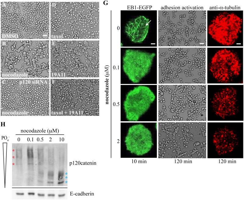
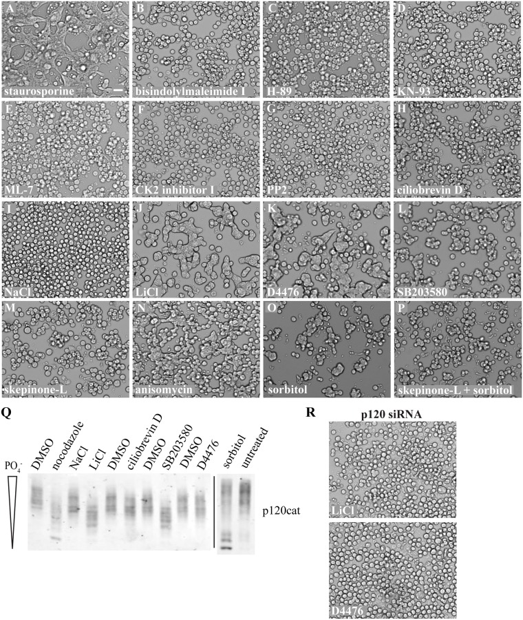
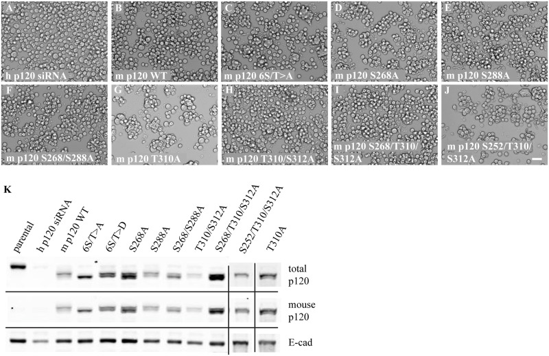
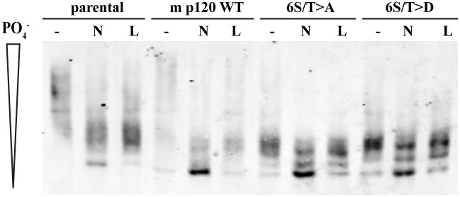
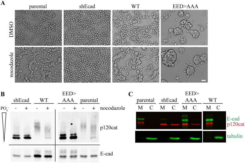
Tight regulation of cadherin-mediated intercellular adhesions is critical to both tissue morphogenesis during development and tissue homeostasis in adults. Cell surface expression of the cadherin-catenin complex is often directly correlated with the level of adhesion, however, examples exist where cadherin appears to be inactive and cells are completely non-adhesive. The state of p120-catenin phosphorylation has been implicated in regulating the adhesive activity of E-cadherin but the mechanism is currently unclear. We have found that destabilization of the microtubule cytoskeleton, independent of microtubule plus-end dynamics, dephosphorylates p120-catenin and activates E-cadherin adhesion in Colo 205 cells. Through chemical screening, we have also identified several kinases as potential regulators of E-cadherin adhesive activity. Analysis of several p120-catenin phosphomutants suggests that gross dephosphorylation of p120-catenin rather than that of specific amino acids may trigger E-cadherin adhesion. Uncoupling p120-catenin binding to E-cadherin at the membrane causes constitutive adhesion in Colo 205 cells, further supporting an inhibitory role of phosphorylated p120-catenin on E-cadherin activity.
Conflict of interest statement
Figures
Similar articles
-
p120-catenin phosphorylation status alters E-cadherin mediated cell adhesion and ability of tumor cells to metastasize.PLoS One. 2020 Jun 26;15(6):e0235337. doi: 10.1371/journal.pone.0235337. eCollection 2020. PLoS One. 2020. PMID: 32589661 Free PMC article.
-
The homeodomain transcription factors Cdx1 and Cdx2 induce E-cadherin adhesion activity by reducing beta- and p120-catenin tyrosine phosphorylation.Am J Physiol Gastrointest Liver Physiol. 2007 Jul;293(1):G54-65. doi: 10.1152/ajpgi.00533.2006. Epub 2007 Apr 26. Am J Physiol Gastrointest Liver Physiol. 2007. PMID: 17463179
-
Conformational epitopes at cadherin calcium-binding sites and p120-catenin phosphorylation regulate cell adhesion.Mol Biol Cell. 2012 Jun;23(11):2092-108. doi: 10.1091/mbc.E11-12-1060. Epub 2012 Apr 18. Mol Biol Cell. 2012. PMID: 22513089 Free PMC article.
-
p120 catenin: an essential regulator of cadherin stability, adhesion-induced signaling, and cancer progression.Prog Mol Biol Transl Sci. 2013;116:409-32. doi: 10.1016/B978-0-12-394311-8.00018-2. Prog Mol Biol Transl Sci. 2013. PMID: 23481205 Free PMC article. Review.
-
Role of p120-catenin in cadherin trafficking.Biochim Biophys Acta. 2007 Jan;1773(1):8-16. doi: 10.1016/j.bbamcr.2006.07.005. Epub 2006 Jul 21. Biochim Biophys Acta. 2007. PMID: 16949165 Review.
Cited by
-
Exposing a novel genetic interaction between unc-33/CRMP and hmp-2/β-catenin during Caenorhabditis elegans embryogenesis.MicroPubl Biol. 2020 Jul 30;2020:10.17912/micropub.biology.000286. doi: 10.17912/micropub.biology.000286. MicroPubl Biol. 2020. PMID: 32760883 Free PMC article. No abstract available.
-
Pharmacological and phosphoproteomic approaches to roles of protein kinase C in kappa opioid receptor-mediated effects in mice.Neuropharmacology. 2020 Dec 15;181:108324. doi: 10.1016/j.neuropharm.2020.108324. Epub 2020 Sep 22. Neuropharmacology. 2020. PMID: 32976891 Free PMC article.
-
E-cadherin in contact inhibition and cancer.Oncogene. 2018 Aug;37(35):4769-4780. doi: 10.1038/s41388-018-0304-2. Epub 2018 May 21. Oncogene. 2018. PMID: 29780167 Free PMC article. Review.
-
Potential Mechanism and Involvement of p120-Catenin in the Malignant Biology of Glioma.J Korean Neurosurg Soc. 2024 Nov;67(6):609-621. doi: 10.3340/jkns.2024.0053. Epub 2024 Jul 3. J Korean Neurosurg Soc. 2024. PMID: 38956806 Free PMC article.
-
Roles for E-cadherin cell surface regulation in cancer.Mol Biol Cell. 2016 Nov 1;27(21):3233-3244. doi: 10.1091/mbc.E16-01-0058. Epub 2016 Aug 31. Mol Biol Cell. 2016. PMID: 27582386 Free PMC article.
References
Publication types
MeSH terms
Substances
Grants and funding
LinkOut - more resources
Full Text Sources
Other Literature Sources
Molecular Biology Databases
Research Materials
Miscellaneous
