. 2016 Feb 4;12(2):e1005822.
doi: 10.1371/journal.pgen.1005822. eCollection 2016 Feb.

Regulation of Stem Cell Proliferation and Cell Fate Specification by Wingless/Wnt Signaling Gradients Enriched at Adult Intestinal Compartment Boundaries

Affiliations

Regulation of Stem Cell Proliferation and Cell Fate Specification by Wingless/Wnt Signaling Gradients Enriched at Adult Intestinal Compartment Boundaries

Ai Tian et al. PLoS Genet. .

Abstract

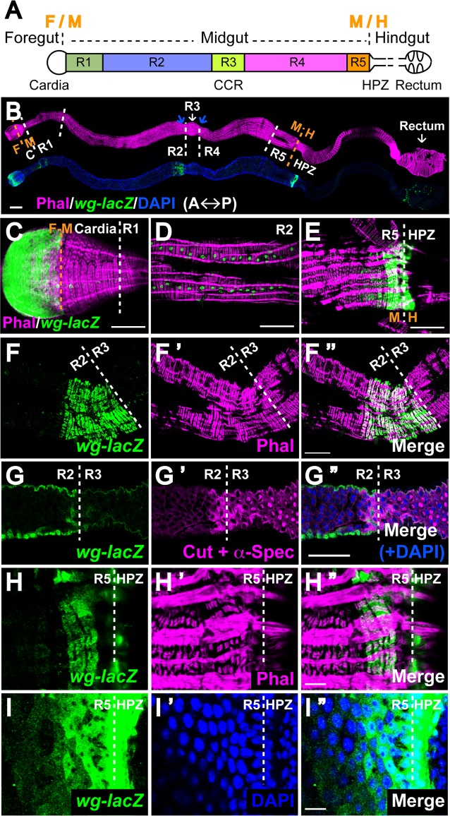
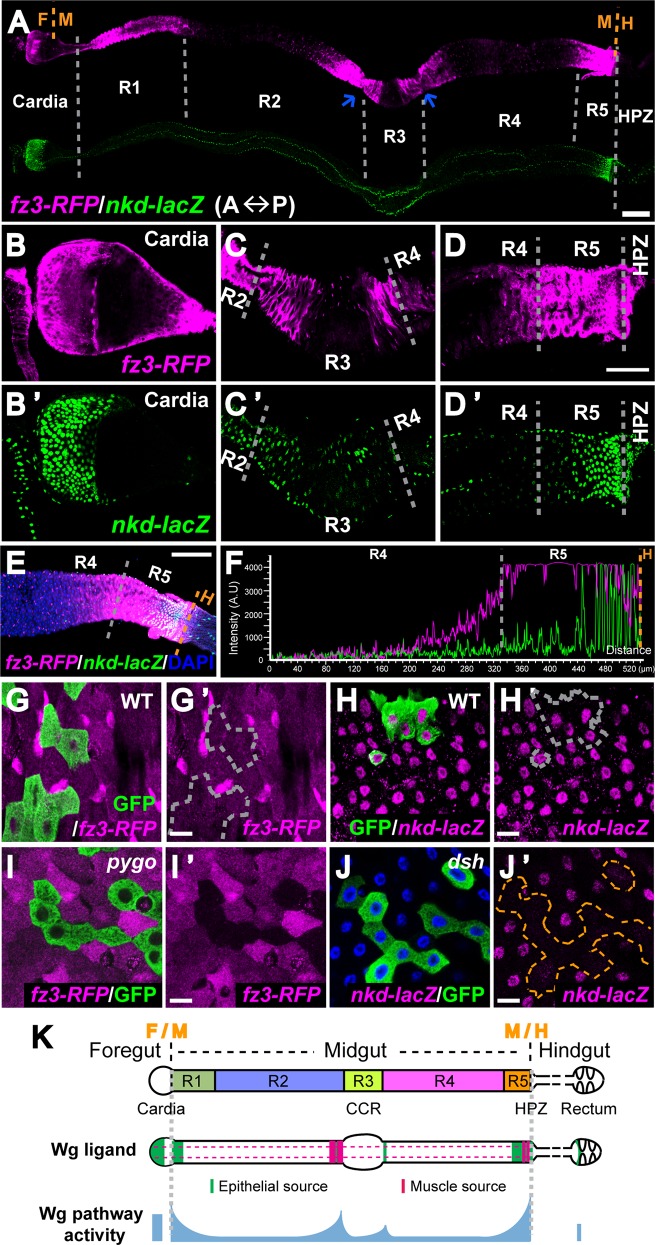
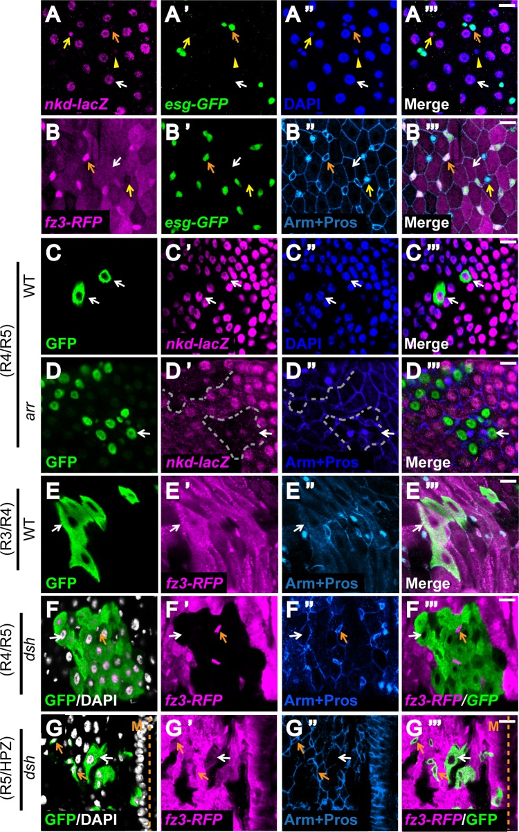
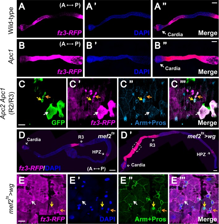
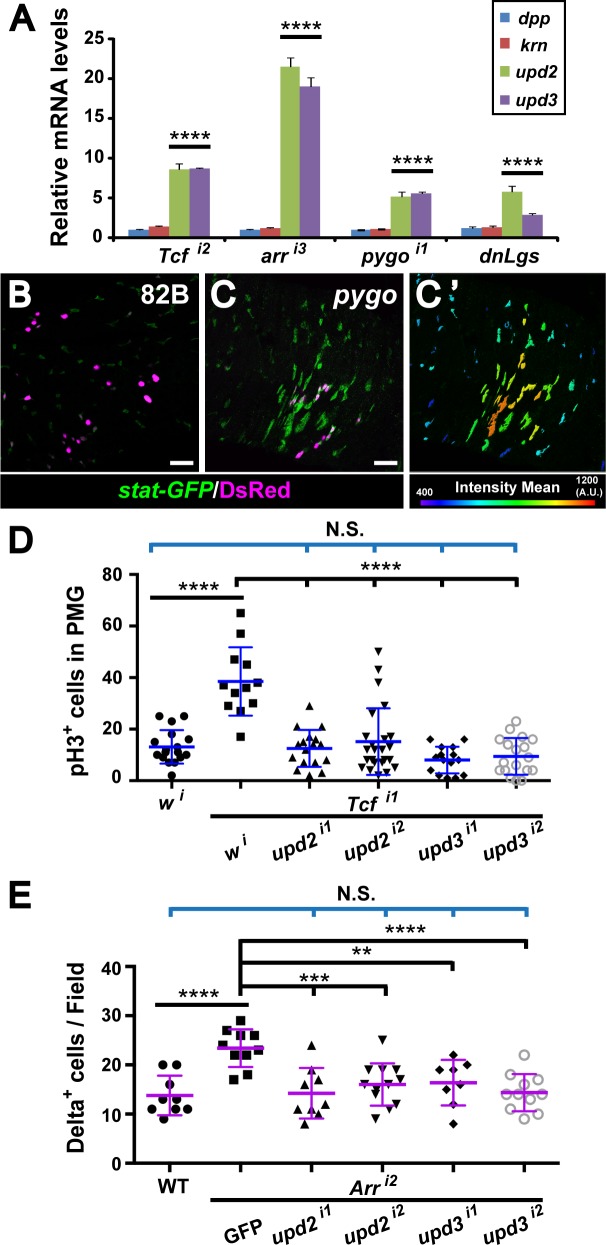
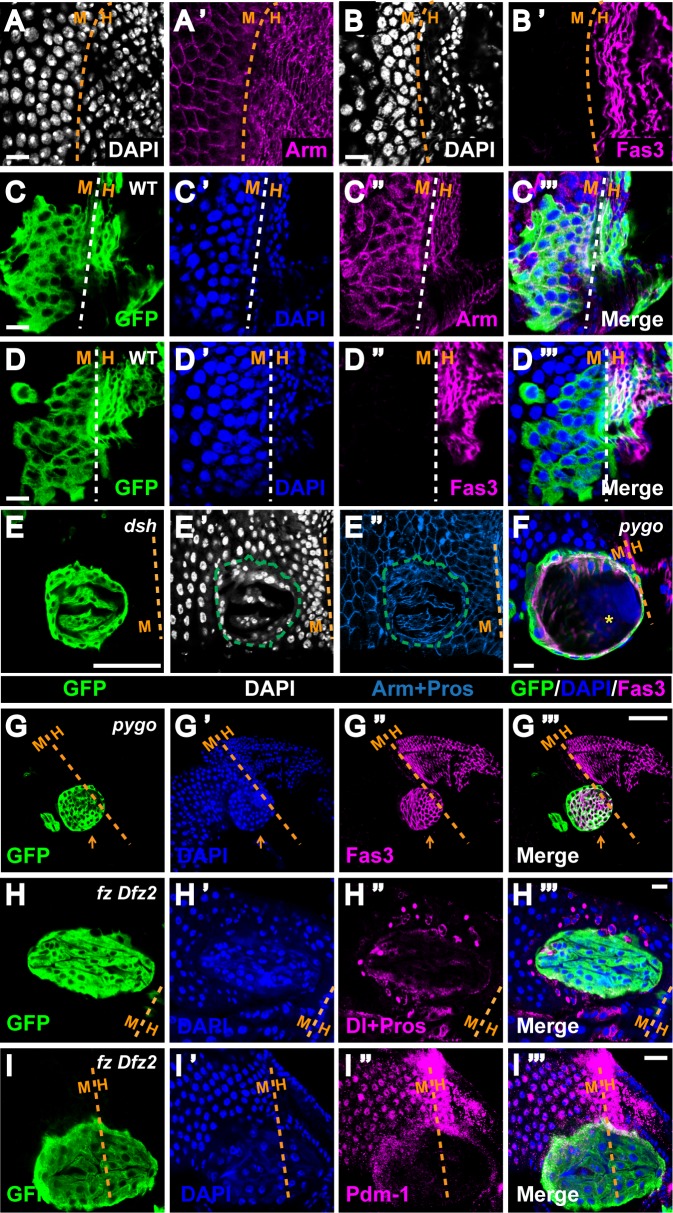
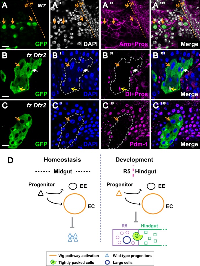
Intestinal stem cell (ISC) self-renewal and proliferation are directed by Wnt/β-catenin signaling in mammals, whereas aberrant Wnt pathway activation in ISCs triggers the development of human colorectal carcinoma. Herein, we have utilized the Drosophila midgut, a powerful model for ISC regulation, to elucidate the mechanisms by which Wingless (Wg)/Wnt regulates intestinal homeostasis and development. We provide evidence that the Wg signaling pathway, activation of which peaks at each of the major compartment boundaries of the adult intestine, has essential functions. Wg pathway activation in the intestinal epithelium is required not only to specify cell fate near compartment boundaries during development, but also to control ISC proliferation within compartments during homeostasis. Further, in contrast with the previous focus on Wg pathway activation within ISCs, we demonstrate that the primary mechanism by which Wg signaling regulates ISC proliferation during homeostasis is non-autonomous. Activation of the Wg pathway in absorptive enterocytes is required to suppress JAK-STAT signaling in neighboring ISCs, and thereby their proliferation. We conclude that Wg signaling gradients have essential roles during homeostasis and development of the adult intestine, non-autonomously controlling stem cell proliferation inside compartments, and autonomously specifying cell fate near compartment boundaries.


Conflict of interest statement

The authors have declared that no competing interests exist.

Figures

Fig 1
Fig 1. Novel sources of Wg in the epithelium and surrounding muscle at adult intestinal compartment boundaries.
(A) Schematic view of the Drosophila adult intestine. It is divided into foregut, midgut and hindgut. The midgut is further demarcated into 5 main compartments, R1-R5. Cardia and rectum are the anterior and posterior ends of the fly gut. CCR: copper cell region, shares the anterior border with R3. HPZ: Hindgut proliferation zone, stands for the most anterior rows of cells of hindgut. (B) Wg expression pattern along the adult gut. Intestines expressing wg-lacZ stained with phalloidin, which marks the muscle layer. Anterior to the left. The blue arrows point to the two major constrictions that denote the position of R3. Scale bar: 100μm. (C-E) Wg-lacZ expression inside cardia, midgut visceral muscles and HPZ. Scale bar: 50μm. (F-G”) Wg-enriched muscle segment anterior to the R2-R3 boundary (marked by the white line). Phalloidin staining reveals the muscle fiber pattern. The appearance of Cutpos α-SpectrinStrong cells delineates the anterior border of R3 as well as CCR. Scale bar: 50μm. (H-I”) Novel muscular and epithelial Wg sources in the terminal midgut (R5) anterior to the R5-HPZ border. Phalloidin marks the muscle layer while DAPI labels the epithelial cell nuclei. Scale bar: 10μm.
Fig 2
Fig 2. Wg pathway activity gradients around adult intestinal compartment boundaries.
(A) Expression pattern of fz3-RFP and nkd-lacZ, two Wg activity reporters, in the adult gut. Anterior to the left. The two blue arrows indicate constriction sites around R3. Scale bar: 100μm. (B-D’) Higher magnification view of (A) around cardia (B and B’), CCR (C and C’) and R5-HPZ region (D and D’). Scale bar: 50μm. Note: the expression of nkd-lacZ around R2-R3 boundary is variable between guts. (E) Merged expression pattern of fz3-RFP and nkd-lacZ from the middle of R4 to the R5-HPZ boundary. Anterior to the left. Scale bar: 100μm. (F) Quantification of fluorescent intensity in (E) from R4 to the R5-HPZ boundary shows gradient expression of both reporters, high at the boundary and low inside compartment. (G-J’) Low-level Wg pathway activities are present inside midgut compartments. Scale bar: 10μm. (G-H’) Wild-type MARCM clones do not affect fz3-RFP or nkd-lacZ expression inside R4. (I and I’) MARCM clones of pygo deplete fz3-RFP signal inside R4. (J and J’) MARCM clones of dsh diminish nkd-lacZ signal inside R4. (K) Schematic view of wg expression and Wg pathway activation in the adult intestine.
Fig 3
Fig 3. Wg pathway is activated primarily in ECs, but not ISCs, during adult homeostasis.
(A-B”‘) Nkd-lacZ and fz3-RFP are expressed in partially distinct cell types. DAPI labels the nuclei and indicates their ploidy. Small Escargotpos Prosperoneg diploid cells are progenitor cells (orange arrow), big polyploid cells are enterocytes (white arrow), small Escargotneg Prosperopos diploid cells are enteroendocrine cells (yellow arrow and arrowhead). (A-A”‘) Nkd-lacZ is primarily expressed inside enterocytes (white arrow) and a subpopulation of enteroendocrine cells (yellow arrow). Scale bar: 10μm. (B-B”‘) Fz3-RFP is expressed in both enterocytes (white arrow) and progenitors (orange arrow). Its expression is mostly absent in the enteroendocrine cells (yellow arrow). Scale bar: 10μm. (C-G”‘) Wg signaling is primarily transduced in enterocytes of the adult gut. MARCM clones of wild-type controls and Wg pathway mutants were induced during larval development and examined soon after eclosion. GFP marks the clones. nkd-lacZ or fz3-RFP represents Wg pathway activity. Orange arrows indicate progenitor cells while white arrows point to enterocytes. (C-D”‘) Nkd-lacZ expression is not affected in wild-type clones (C-C”‘) but specifically lost within enterocytes of arr clones at intestinal compartment boundaries (D-D”‘). (E-F”‘) Fz3-RFP expression is not affected in wild-type clones (E-E”‘) but specifically lost within enterocytes of dsh clones at intestinal compartment boundaries (F-F”‘). The mutant enterocytes retain normal cell morphology, nuclear morphology and cell-cell junctions, indicating that the loss of Wg pathway activation in this cell type is not due to cell death. Note that Fz3-RFP expression in progenitors is not dependent on Wg signaling. (G-G”‘) At the unique R5-HPZ boundary, Wg pathway is activated in both progenitors and enterocytes. Scale bar: 10μm.
Fig 4
Fig 4. All gut cell types are capable of responding to Wg exposure.
(A-B”) Compared with the wild-type anterior midgut, Wg signaling is ectopically induced in the Apc1Q8 mutant. Fz3-RFP serves as a reporter for Wg pathway activity. DAPI labels the gut cell nuclei. Anterior to the left. Scale bar: 100μm. (C-C”‘) GFP-marked Apc2 Apc1 mutant MARCM clones exhibit aberrantly high fz3-RFP signals in all gut cell types at compartment boundaries, including progenitors (orange arrow), enterocytes (white arrow) and enteroendocrine cells (yellow arrow), as indicated by Arm and Prospero staining. Of note, despite the high-level Wg pathway activation that is already present at compartment boundaries, Apc2 Apc1 double mutant cells display even higher levels of activation at all boundaries. Scale bar: 10μm. (D and D’) Wg originating from the visceral muscle is sufficient to activate signaling in the intestinal epithelium. Wg is expressed throughout the muscle using a temperature sensitive dMef2-Gal4 driver (D’) alongside with wild-type control (D). Flies were shifted to the permissive temperature for wg expression at eclosion and reared at this temperature for one week prior to analysis. Dramatic induction of fz3-RFP is detected in the epithelium, most pronouncedly in the anterior midgut. Scale bar: 100μm. (E-E”‘) Overexpression of wg in muscle during adulthood induces ectopic Wg pathway activation in all gut cell types, including progenitors (orange arrow), enterocytes (white arrow) and enteroendocrine cells (yellow arrow), as indicated by Arm and Prospero staining. Scale bar: 10μm.
Fig 5
Fig 5. Disruption of Wg signaling non-autonomously induces proliferation of ISCs.
(A) MARCM clones were induced on the day of eclosion and analyzed 5–7 days later. Quantification of clone size for wild-type, pygo and dsh ISC lineages in the posterior midgut shows that both pygo and dsh have a higher percentage of multi-cellular clones compared with wild-type. Number of clones examined: 82B (n = 864), pygo (n = 898), 19A (n = 384) and dsh (n = 699). (B) Wild-type progenitor cells, marked with esg-lacZ, aberrantly gather around the adult pygo mutant MARCM clones (marked by GFP, inside R4). Scale bar: 25μm. (C-F) The abnormally clustered wild-type progenitor cells include both ISCs and EBs. Scale bar: 10μm. (C and D) Increased number of ISCs, marked by Dl-lacZ, are aberrantly clustered around arr MARCM clones (inside R4) (D), this defect was not observed when wild-type clones were induced (C). (E and F) Increased number of EBs, marked by Su(H)-lacZ, are aberrantly clustered around pygo MARCM clones (inside R4) (F), this defect was not observed when wild-type clones were induced (E). (G) Phospho-histone H3 labels cells that are undergoing mitosis and serves as a marker for proliferation. Quantification of pH3+ cells in posterior midguts bearing either pygo or dsh MARCM clones, alongside wild-type controls, reveals a significant increase in proliferation. Number of guts examined: 82B (n = 15), pygo (n = 21), 19A (n = 20) and dsh (n = 19). ****P<0.0001 (t-test). (H) Reducing Wg pathway activity specifically in adult enterocytes using RNAis (i, the number after i indicates the line used) also results in increased proliferation. Number of guts examined: wild-type (n = 15), TCFi1 (n = 17), arri1 (n = 16) and pygoi1 (n = 17). ****P<0.0001; ***P<0.001 (t-test). (I-L) Diminishing Wg pathway activity specifically in adult enterocytes also results in disorganized gut epithelium (J-L). Wild-type control is shown in (I). Scale bar: 10μm.
Fig 6
Fig 6. JAK-STAT pathway overactivation upon disruption of Wg signaling in enterocytes.
(A) When the MyoIAts driver was used to knockdown Wg signaling inside enterocytes during adulthood, mRNA expression levels of upd2 and upd3, but not dpp or krn, are greatly induced. The qPCR results were normalized to rpl32 and compared with wild-type, presented as mean fold-change with standard deviation. ****P<0.0001 (t-test). (B-C’) JAK-STAT pathway activity is abruptly increased in the vicinity of the adult pygo mutant MARCM clones (inside R4). Stat-GFP serves as a reporter for JAK/STAT signaling. The pygo clones are marked with DsRed. Scale bar: 50μm. (C’) Intensity of stat-GFP expression is quantified using the IMARIS software. (D and E) JAK-STAT pathway activation mediates the non-autonomous effects of Wg pathway disruption. (D) Knockdown of TCF by RNAi inside enterocytes using the MyoIAts driver results in ISC overproliferation, and this could be suppressed by simultaneous inactivation of JAK-STAT pathway via upd2 or upd3 knockdown. Number of guts examined: wi (n = 17), TCFi1+ wi (n = 12), TCFi1+ upd2i1 (n = 17), TCFi1+ upd2i2 (n = 24), TCFi1+ upd3i1 (n = 15) and TCFi1+ upd3i2 (n = 18). ****P<0.0001; N.S. not significant (t-test). (E) Knockdown arr by RNAi inside enterocytes using the MyoIAts driver results in increase of Deltapos cells, which could also be suppressed by further knockdown of JAK-STAT pathway activity. Number of guts examined: wild-type (n = 9), Arri2+ GFP (n = 10), Arri2+ upd2i1 (n = 9), Arri2+ upd2i2 (n = 12), Arri2+ upd3i1 (n = 8) and Arri2+ upd3i2 (n = 11). ****P<0.0001; ***P<0.001; **P<0.01; N.S. not significant (t-test).
Fig 7
Fig 7. Disruption of Wg signaling at R5-HPZ boundary induces masses of “tightly-packed” cells.
(A-B’) Terminal midgut cells and HPZ hindgut cells separated by the midgut-hindgut boundary are derived from distinct origins (endoderm for midgut versus ectoderm for hindgut). They have disparate nuclei shape, nuclei size and cell size (A and B), as well as disparate cell-cell junctions (A’ and B’). Dashed lines mark the midgut-hindgut border with midgut on the left. DAPI labels the nuclei. In R5, cells are large and have round nuclei, whereas the HPZ is composed of tightly-spaced rows of small, columnar cells. Arm and Fas3 demarcate the adherence junctions. Note that Fas3 is expressed at high level in the hindgut and its expression drops dramatically in the midgut (B’). Scale bar: 10μm. (C-D”‘) Wild-type MARCM clones crossing the midgut-hindgut boundary do not affect nuclei morphology or cell-cell junction on either side. The border between the two compartments is clearly demarcated. Note that Fas3 is restricted to the hindgut side. Scale bar: 10μm. (E-I”‘) MARCM clones of indicated mutants were induced during larval development and examined soon after eclosion. GFP marks the clones. Dashed lines mark the midgut-hindgut border with midgut on the left. (E-E”‘) A tightly packed dsh clone exhibits abnormal spherical arrangement of nuclei (DAPI) as well as distorted cellular organization (Arm). Scale bar: 10μm. (F) A tightly packed pygo clone extends outside the gut. Cross-section of the clone shows that it is hollow with a haze of DAPI inside (yellow asterisk). Note that high-level Fas3 signal is localized at the inner surface of the pygo clone, and is absent from its outline, suggesting that cells within the clone has maintained apico-basal polarity. Scale bar: 10μm. (G-G”‘) A tightly packed pygo clone (orange arrow) expresses Fas3 at a high level similar to that of the hindgut despite its location inside the midgut. Again, high-level Fas3 signal is localized at the inner surface of the pygo clone, and is absent from its outline. In addition, the clone has extended outside the gut outline. Scale bar: 50μm. (H-H”‘) A tightly packed fz Dfz2 clone is devoid of Deltapos and Prosperopos cells except at the very periphery. Scale bar: 10μm. (I-I”‘) A tightly packed fz Dfz2 clone is devoid of Pdm-1pos cells. Scale bar: 10μm.
Fig 8
Fig 8. Disruption of Wg signaling at R5-HPZ boundary induces “large” cells.
(A-C”‘) MARCM clones of Wg pathway components were induced during larval development and examined soon after eclosion. GFP marks the clones. Dashed lines mark the midgut-hindgut border with midgut on the left. (A-A”‘) Disrupting Wg pathway activity by arr clones results in cells with dramatically larger nuclei and cell size as well as distorted cell outlines (orange arrows). Scale bar: 10μm. (B-B”‘) The mutant fz Dfz2 clones bearing large cells (orange arrows) have normal looking Deltapos ISCs (white arrow) and Prosperopos EEs (yellow arrow). Scale bar: 10μm. (C-C”) The mutant fz Dfz2 clones bearing large cells (orange arrows) have greatly diminished Pdm-1 staining. Scale bar: 10μm. (D) Model: roles of Wg signaling in the fly gut under normal conditions. (Left) During adult homeostasis, proper Wg pathway activation inside the enterocytes non-autonomously prevents ISC over-proliferation. (Right) At the midgut-hindgut boundary, Wg is transduced both in progenitors and enterocytes. During development, normal Wg signaling around this boundary is required for cell fate specification and prevention of lineage mixing.

Similar articles

Cited by

References

    1. MacDonald BT, Tamai K, He X (2009) Wnt/beta-catenin signaling: components, mechanisms, and diseases. Dev Cell 17: 9–26. 10.1016/j.devcel.2009.06.016 - DOI - PMC - PubMed
    1. Clevers H, Nusse R (2012) Wnt/beta-catenin signaling and disease. Cell 149: 1192–1205. 10.1016/j.cell.2012.05.012 - DOI - PubMed
    1. Brunner E, Peter O, Schweizer L, Basler K (1997) pangolin encodes a Lef-1 homologue that acts downstream of Armadillo to transduce the Wingless signal in Drosophila. Nature 385: 829–833. - PubMed
    1. van de Wetering M, Cavallo R, Dooijes D, van Beest M, van Es J, et al. (1997) Armadillo coactivates transcription driven by the product of the Drosophila segment polarity gene dTCF. Cell 88: 789–799. - PubMed
    1. Kramps T, Peter O, Brunner E, Nellen D, Froesch B, et al. (2002) Wnt/wingless signaling requires BCL9/legless-mediated recruitment of pygopus to the nuclear beta-catenin-TCF complex. Cell 109: 47–60. - PubMed

Publication types

MeSH terms

LinkOut - more resources