. 2016 Feb 4;11(2):e0148083.
doi: 10.1371/journal.pone.0148083. eCollection 2016.

Extracellular Vesicles from BOEC in In Vitro Embryo Development and Quality

Affiliations

Extracellular Vesicles from BOEC in In Vitro Embryo Development and Quality

Ricaurte Lopera-Vásquez et al. PLoS One. .

Abstract

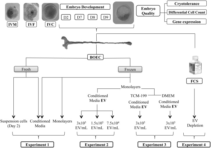
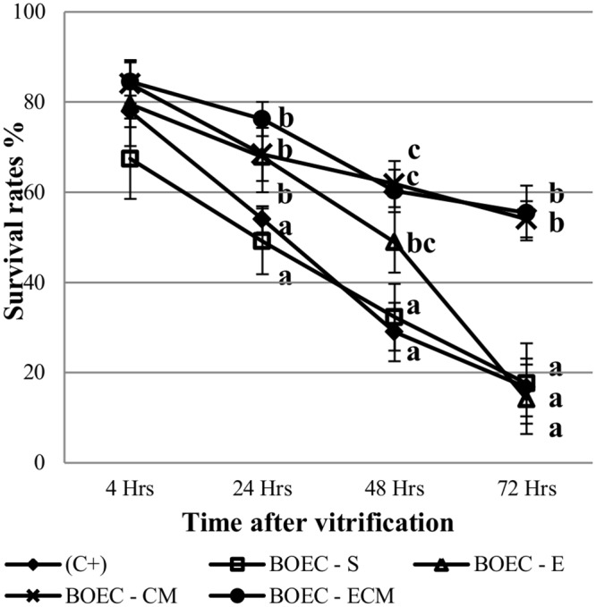
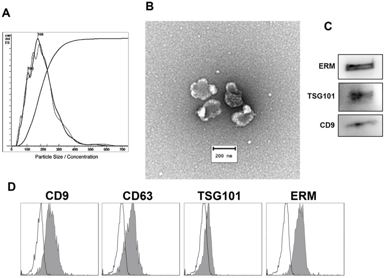
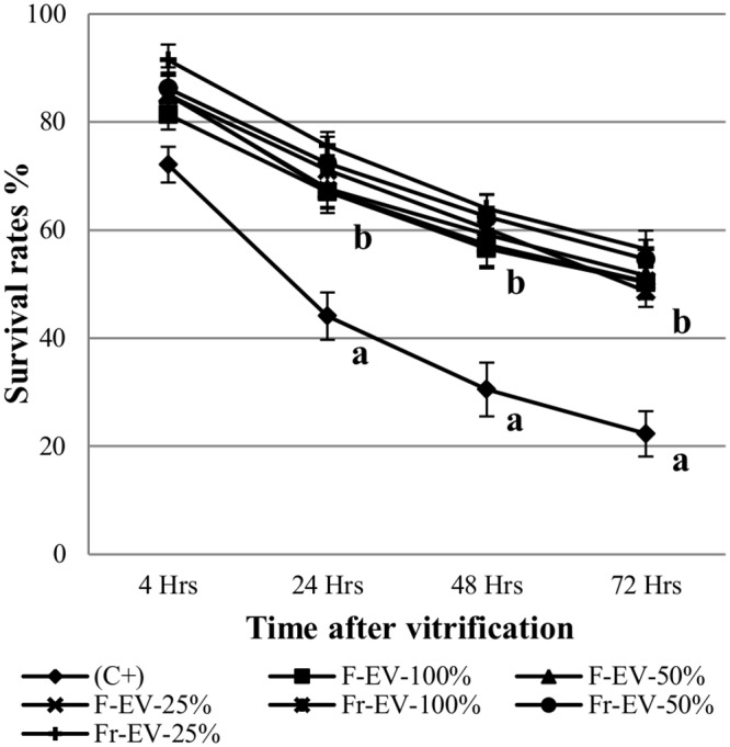
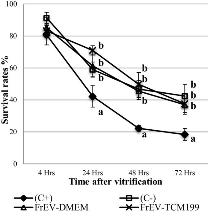
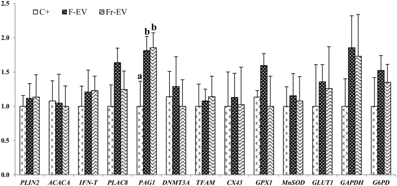
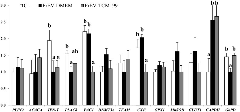
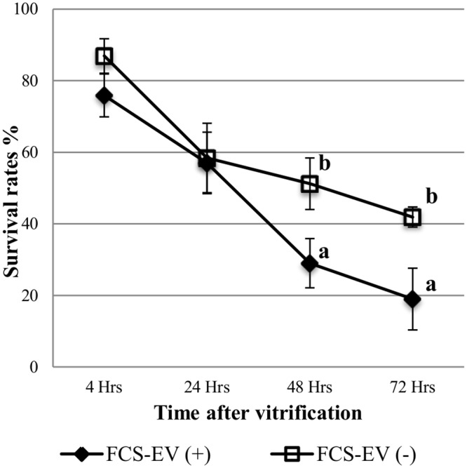
To evaluate the effect of conditioned media (CM) and Extracellular Vesicles (EVs) derived from bovine oviduct epithelial cell (BOEC) lines on the developmental capacity of bovine zygotes and the quality of embryos produced in vitro, presumptive zygotes were cultured under specific conditions. In experiment 1, zygotes were cultured either on monolayers from BOEC extended culture (E), together with fresh BOEC suspension cells, or with BOEC-CM from fresh or E-monolayers. In experiment 2, EVs were isolated from BOEC-CM and characterized (150-200 nm) by Nanosight® and electron microscopy. Zygotes were cultured in the presence of 3x10(5) EVs/mL, 1.5x10(5) EVs/mL or 7.5x10(4) EVs/mL of fresh or frozen BOEC-EVs. In experiment 3, zygotes were cultured in absence of FCS but with EVs from BOEC-E that had been cultured in different culture media. In experiment 4, zygotes were cultured in SOF+5% normal-FCS, or EV-depleted-FCS. In all cases, cleavage rate (Day 2) and blastocyst development (Day 7-9) was assessed. Blastocysts on Days 7/8 were used for quality evaluation through differential cell count, cryotolerance and gene expression patterns. No differences were found among all FCS-containing groups in cleavage rate or blastocyst yield. However, embryos derived from BOEC-CM had more trophectoderm cells, while embryos derived from BOEC-EVs, both fresh and frozen, has more trophectoderm and total cells. More embryos survived vitrification in the BOEC-CM and BOEC-EV groups. In contrast, more embryos survived in the EV-depleted-FCS than in normal-FCS group. Gene expression patterns were modified for PAG1 for embryos cultured with EVs in the presence of FCS and for IFN-T, PLAC8, PAG1, CX43, and GAPDH in the absence of FCS. In conclusion, EVs from FCS have a deleterious effect on embryo quality. BOEC-CM and EVs during in vitro culture had a positive effect on the quality of in vitro produced bovine embryos, suggesting that EVs have functional communication between the oviduct and the embryo in the early stages of development.


Conflict of interest statement

Competing Interests: The authors have declared that no competing interests exist.

Figures

Fig 1
Fig 1. Experimental design.
Fig 2
Fig 2. Survival rate after vitrification and warming of D7 blastocysts co-cultured with different types of BOEC (Suspension Cells: BOEC-S [n = 69], Extended Culture: BOEC-E [n = 53]) or Conditioned Media (from fresh: BOEC-CM [n = 70], or extended culture monolayers-BOEC-ECM [n = 66]) and Control+FCS (C+; n = 113).
a,b,cDifferent superscripts indicate significant differences at given time points (p<0.05).
Fig 3
Fig 3. Characterization of vesicles isolated from BOEC-CM.
A- Nanoparticle tracking analysis (NTA) of a representative EV sample. B- Transmission electron microscope image of negative-stained BOEC-EVs. C- Western-blot analysis of BOEC-EV lysates with EV markers. D- Bead-assisted flow cytometry analysis of EV isolated from BOEC-ECM. EV-coupled beads were stained for CD9, CD63, TSG101 and ERM EV markers. Negative control is depicted as an empty plot.
Fig 4
Fig 4. Survival rate after vitrification and warming of Day 7 blastocysts cultured with different concentrations (100, 50, 25%) of recently purified (F-EV; n = 96, 110, 94 respectively) or frozen/thawed (Fr-EV; n = 83, 86, 87 respectively)) BOEC extracellular vesicles.
a,bDifferent superscripts indicate significant differences at given time points (p<0.05).
Fig 5
Fig 5. Survival rate after vitrification and warming of D7-8 blastocysts cultured with extracellular vesicles (EV) secreted by BOEC cultured in DMEM (n = 61), TCM199 (n = 59) or in Control+FCS (C+; n = 46) and C-: Control-FCS (C+; n = 50).
a,b,Different superscripts indicate significant differences at given time points (p<0.05).
Fig 6
Fig 6. Relative mRNA transcription in bovine in vitro blastocysts (D7 p.i) cultured with or without (C+) EV secreted by BOEC fresh (F-EV) and frozen (Fr-EV) of genes related with fatty acid metabolism (PLIN2, ACACA), implantation (IFN-T, PLAC8, PAG1), epigenetics (DNMT3A, TFAM), gap junctions (CX43) and oxidative stress (GPX1,MNSOD, GLUT1, GAPDH, G6PD).
Data are relative to mean of internal genes H2AFZ, and 18S. Data presented are mean±SE. a.b Different superscripts indicate significant differences for each gene (p<0.05).
Fig 7
Fig 7. Relative mRNA transcription in bovine in vitro blastocysts (D7-8 p.i) cultured with or without (C-) EV secreted by BOEC cultured in DMEM and TCM199 of genes related with fatty acid metabolism (PLIN2, ACACA), implantation (IFN-T, PLAC8, PAG1), epigenetics (DNMT3A, TFAM), gap junctions (CX43) and oxidative stress (GPX1,MNSOD, GLUT1, GAPDH, G6PD).
Data are relative to mean of internal genes H2AFZ, and 18S. Data presented are mean±SE. a.b Different superscripts indicate significant differences for each gene (p<0.05).
Fig 8
Fig 8. Survival rate after vitrification and warming of D7 blastocysts cultured with normal FCS (containing EV; n = 46) or EV-depleted FCS; n = 49.
a,bDifferent superscripts indicate significant differences at given time points (p<0.05).

References

    1. Rizos D, Clemente M, Bermejo-Alvarez P, De La Fuente J, Lonergan P, Gutiérrez-Adán A. Consequences of In Vitro Culture Conditions on Embryo Development and Quality. Reprod Domest Anim. 2008;43: 44–50. 10.1111/j.1439-0531.2008.01230.x - DOI - PubMed
    1. Plourde D, Vigneault C, Laflamme I, Blondin P, Robert C. Cellular and molecular characterization of the impact of laboratory setup on bovine in vitro embryo production. Theriogenology. 2012;77: 1767–1778.e1. 10.1016/j.theriogenology.2011.12.021 - DOI - PubMed
    1. Niemann H, Wrenzycki C. Alterations of expression of developmentally important genes in preimplantation bovine embryos by in vitro culture conditions: implications for subsequent development. Theriogenology. 2000;53: 21–34. - PubMed
    1. Pontes JHF, Nonato-Junior I, Sanches BV, Ereno-Junior JC, Uvo S, Barreiros TRR, et al. Comparison of embryo yield and pregnancy rate between in vivo and in vitro methods in the same Nelore (Bos indicus) donor cows. Theriogenology. 2009;71: 690–697. 10.1016/j.theriogenology.2008.09.031 - DOI - PubMed
    1. Hackett AJ, Durnford R, Mapletoft RJ, Marcus GJ. Location and status of embryos in the genital tract of superovulated cows 4 to 6 days after insemination. Theriogenology. 1993;40: 1147–1153. 10.1016/0093-691X(93)90285-D - DOI

Publication types