. 2016 Jun;63(6):1987-2003.
doi: 10.1002/hep.28486. Epub 2016 Mar 15.

c-Jun N-terminal kinase mediates mouse liver injury through a novel Sab (SH3BP5)-dependent pathway leading to inactivation of intramitochondrial Src

Affiliations

c-Jun N-terminal kinase mediates mouse liver injury through a novel Sab (SH3BP5)-dependent pathway leading to inactivation of intramitochondrial Src

Sanda Win et al. Hepatology. 2016 Jun.

Abstract

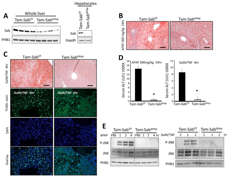
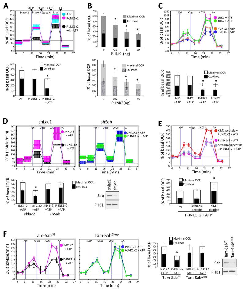
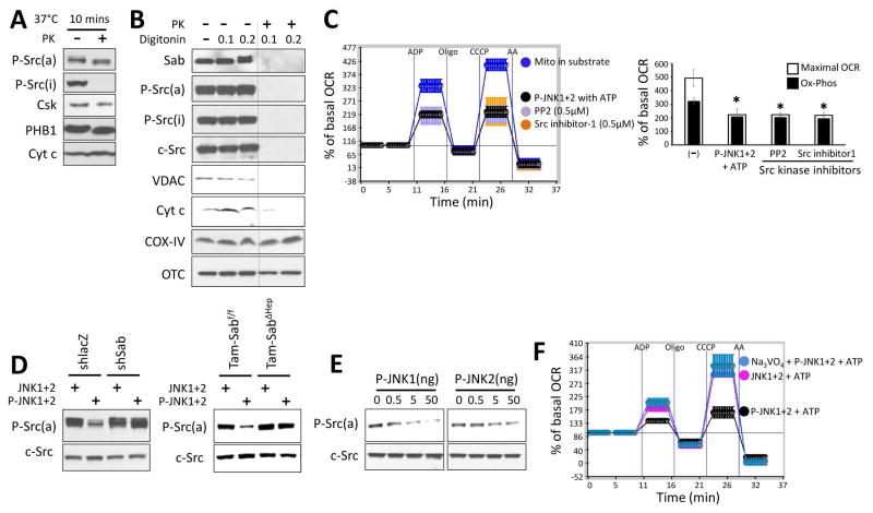
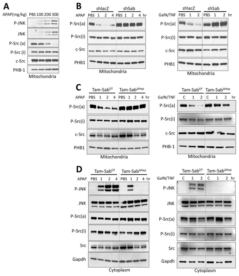
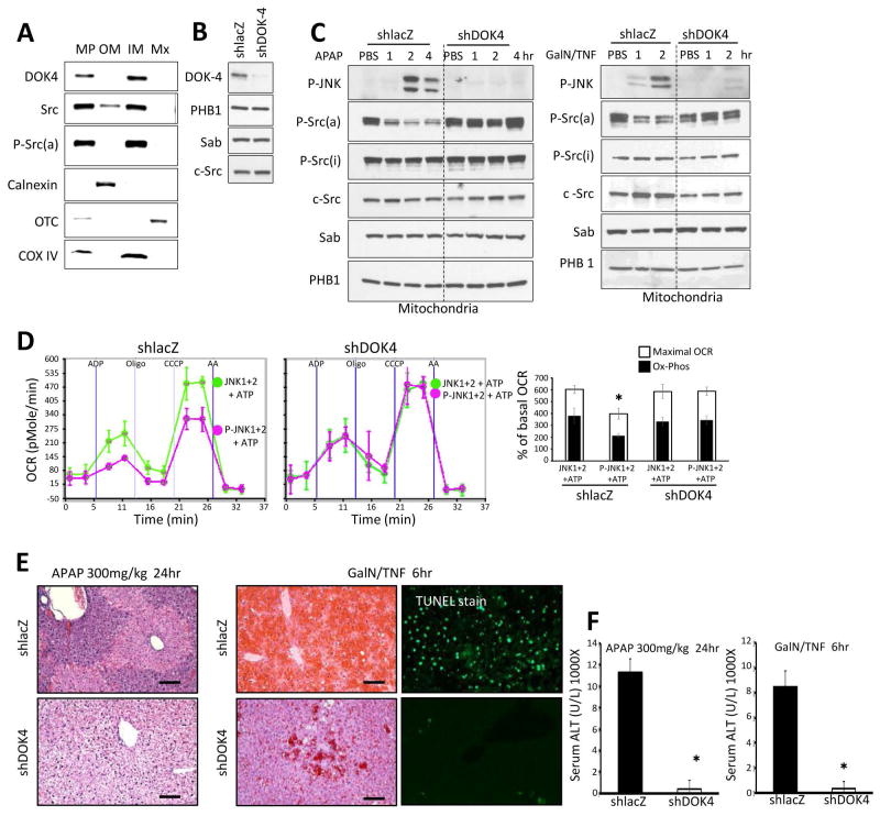
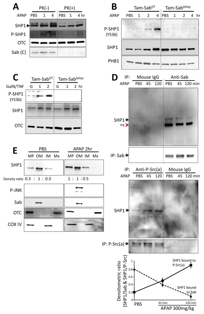
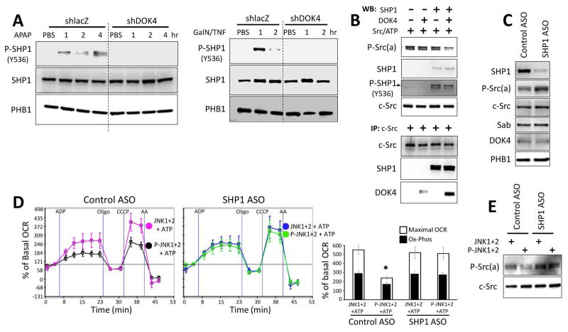
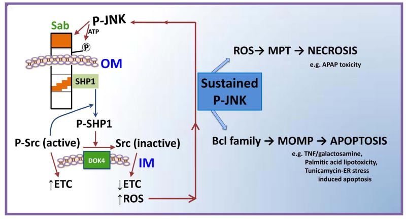
Sustained c-Jun N-terminal kinase (JNK) activation has been implicated in many models of cell death and tissue injury. Phosphorylated JNK (p-JNK) interacts with the mitochondrial outer membrane SH3 homology associated BTK binding protein (Sab, or SH3BP5). Using knockdown or liver-specific deletion of Sab, we aimed to elucidate the consequences of this interaction on mitochondrial function in isolated mitochondria and liver injury models in vivo. Respiration in isolated mitochondria was directly inhibited by p-JNK + adenosine triphosphate. Knockdown or liver-specific knockout of Sab abrogated this effect and markedly inhibited sustained JNK activation and liver injury from acetaminophen or tumor necrosis factor/galactosamine. We then elucidated an intramitochondrial pathway in which interaction of JNK and Sab on the outside of the mitochondria released protein tyrosine phosphatase, nonreceptor type 6 (SHP1, or PTPN6) from Sab in the inside of the mitochondrial outer membrane, leading to its activation and transfer to the inner membrane, where it dephosphorylates P-Y419Src (active), which required a platform protein, docking protein 4 (DOK4), on the inner membrane. Knockdown of mitochondrial DOK4 or SHP1 inhibited the inactivation of mitochondrial p-Src and the effect of p-JNK on mitochondria.

Conclusions: The binding to and phosphorylation of Sab by p-JNK on the outer mitochondrial membrane leads to SHP1-dependent and DOK4-dependent inactivation of p-Src on the inner membrane; inactivation of mitochondrial Src inhibits electron transport and increases reactive oxygen species release, which sustains JNK activation and promotes cell death and organ injury. (Hepatology 2016;63:1987-2003).


Conflict of interest statement

Conflict of interest statement: S.W, T.A.T, R.W.M.M and N.K have declared that no conflict of interest exists. M.A. is an employee and shareholder of Ionis Pharmaceuticals.

Figures

Fig. 1
Fig. 1. Effect of conditional knockout of Sab in liver injury models
Mice were fed with tamoxifen diet (Tam-diet) for 7 days. (A) Depletion of Sab in mitochondria isolated from whole liver homogenate with residual Sab presumably in nonparenchymal cells and undetectable Sab in freshly isolated mouse hepatocytes. One week later tamoxifen treated littermate control (Tam-Sabf/f Control) and tamoxifen treated Sabf/f;Alb-CreERT2+/− Sab KO (Tam-SabΔHep KO) were treated with APAP 300mg/kg i.p or TNF 12μg/kg/GalN 800mg/kg i.p. (B) Comparison of H and E liver histology 24 hours after APAP. Scale bars represent 100μm. (C) Comparison of H and E and TUNEL stain of liver 6 hours after TNF/GalN. Scale bars represent 100μm. (D) Serum ALT 24 hours after APAP or 6 hours after TNF/GalN. Error bars represent S.D of 3 separate experiments. N = 5; (*) represent p <0.05 (t-test), Tam-Sabf/f Control vs Tam-SabΔHep KO (E) Comparison of P-JNK translocation to mitochondria in early hours after APAP or GalN/TNF in Tam-Sabf/f versus Tam-SabΔHep mice.
Fig. 2
Fig. 2. Sab dependent effect of P-JNK on OCR of isolated mitochondria
(A) Mitochondrial Oxygen consumption rate (OCR) of isolated mitochondria (20μg) from wild type mouse liver treated with activated JNK (P-JNK1+2 50ng each) with or without ATP (6μM final) in 50μl mitochondrial assay solution supplemented with pyruvate/malate. Data represents real-time continuous measurement of OCR using in Seahorse XF24 analyzer. (*) represent p <0.05 (t-test), vs P-JNK or ATP alone. (B) OCR of isolated mitochondria treated with serially diluted P-JNK1 or P-JNK2 in the presence of ATP and pyruvate/malate. (*) represent p <0.05 (paired t-test) for both mitochondrial maximal oxidative capacity and oxidative phosphorylation, vs without P-JNK. (C) Mitochondria (20μg) were treated with 50ng of recombinant unactivated JNK (JNK1 or JNK2) or activated JNK (P-JNK1 or P-JNK2) in the presence of ATP and pyruvate/malate. (*) represent p <0.05, JNK vs P-JNK. (D) Adenoviral shSab or adenoviral shlacZ was given to mice intravenously and 10 days later the mitochondria were isolated. Efficiency of Sab knockdown was confirmed by western blot of isolated mitochondria. 20μg of mitochondria were treated with unactivated JNK1+2 or P-JNK1+2 (50ng each) in the presence of ATP and pyruvate/malate. (*) represents p <0.05, shlacZ vs shSab treated with P-JNK1+2 + ATP. (E) Mitochondria (20μg) isolated from wild type mice were treated with P-JNK1+2 with scrambled peptide or KIM1 peptide in the presence of ATP. (F) Mitochondria (20μg) isolated from Tam-Sabf/f *ATP and pyruvate/malate. Efficiency of Sab knockout was confirmed by western blot of isolated mitochondria. (*) represents p <0.05, Tam-Sabf/f Control vs Tam-SabΔHep KO treated with P-JNK+ATP. The OCR was measured in following order: after acquisition of basal (State 2) OCR, state 3 respiration after ADP injection, state 4 respiration after oligomycin injection, maximal respiration after CCCP injection, and non-mitochondrial OCR after antimycinA (AA) were measured. Error bars represent the S.D of triplicate or pentaplicate samples in one representative experiment and bar graphs compare maximum respiration and oxidative phosphorylation (OCR after ADP minus oligomycin) for three experiments. In bar graph, (*) indicates both max OCR and oxidative phosphorylation were significant.
Fig. 3
Fig. 3. Localization of Src in mitochondria and effect of P-JNK on P-Src
(A) Isolated mitochondria from wild type mice were incubated with or without proteinase K (PK) (1mg/ml) at 37°C for 10 min. (B) Isolated mitochondria were incubated with digitonin 0.1 mg or 0.2 mg per mg of mitochondria protein in ice for 30min followed by PK (1mg/ml) (or no PK) for 10 minutes at 37°C. Western blot analysis in A and B was performed using antisera against Sab, P-Src(a) (active form, Y419), P-Src(i) (inactive form, Y527), c-Src, VDAC, cytochrome c (Cyt c), cytochrome oxidase (COX) IV and ornithine carbamoyl-transferase (OTC). (C) Mitochondria were incubated with substrate (pyruvate/malate) alone or substrate with P-JNK plus ATP, or PP2 (Src inhibitor), or Src inhibitor 1. Error bars represent the S.D of pentaplicate samples in one representative experiment of 3 different preps summarized in bar graph. (*) represent p <0.05, vs substrate alone. (D) Effect of JNK on Src activity in isolated liver mitochondria. Mitochondria from shlacZ or shSab treated mice or mitochondria from Tam-Sabf/f Control or Tam-SabΔHep KO mice were incubated as in (C) and western blot was performed for P-Src(a) and c-Src. Western blots are representative of 3 different preps. (E) Immunoblots of isolated mitochondria treated with serially diluted P-JNK1 or P-JNK2 in the presence of ATP and pyruvate/malate. (F) Isolated liver mitochondria from wild type mice were incubated in pyruvate/malate with unactivated JNK+ATP or P-JNK+ATP with or without sodium thiovanadate 2mM. Error bars represent the S.D of pentaplicate samples in one representative experiment of 3 different preps.
Fig. 4
Fig. 4. Sab dependence of mitochondrial Src inactivation in liver injury models in vivo
(A) Wild type mice were treated with PBS or APAP 100, 200, 300 mg/kg i.p and mitochondria were isolated at 2 hours later for western blot analysis. (B) Ad-shlacZ or Ad-shSab was given to wild type mice intravenously and 10 days later treatments with APAP (300mg/kg i.p) in warm PBS or D-GalN (800mg/kg i.p)/TNF-α (12μg/kg i.p) in pyrogen-free PBS. D-GalN was given 30 min prior to TNF-α. (C, D) Mice were fed with Tam-diet for 7 days. One week later Tam-Sabf/f Control and Tam-SabΔHep KO were treated with APAP or TNF/GalN. Mitochondria (C) and cytoplasm (post mitochondria) (D) were isolated from liver at indicated time points by differential centrifugation. Western blot analysis was performed using antisera against P-Src(a) (Y419), P-Src(i) (Y527), c-Src, P-JNK, JNK, PHB1 and Gapdh. Western blots are representative of 3 different experiments.
Fig. 5
Fig. 5. DOK4 dependence of JNK effect on isolated mitochondria in vivo and in vitro
(A) Isolated mitochondria were fractionated into mitoplasts (MP), outer membrane (OM), inner membrane (IM) and matrix (MX). Western blot was then performed using marker enzymes to verify fractionation. Note, DOK4 and P-Src(a) were localized to the inner membrane. (B) Ad-shlacZ or Ad-shDOK4 was given to wild type mice intravenously and 10 days later efficiency of DOK4 knockdown was confirmed by western blot of isolated mitochondria. (C) Mice were treated with APAP (300mg/kg i.p) in warm PBS or D-GalN (800mg/kg i.p)/TNF-α (12μg/kg i.p) in pyrogen-free PBS. D-GalN was given 30min prior to TNF-α. Mitochondria were isolated at indicated times for western blot analysis using DOK4, P-JNK, P-Src(a), P-Src(i), c-Src, Sab and PHB1. Western blots are representative of 3 different preps. (D) Mitochondria (20μg) isolated from shlacZ or shDOK4 treated mice were incubated with pyruvate/malate and treated with JNK1+2 or P-JNK1+2 in the presence of ATP. Error bars in E represent the S.D of pentaplicate of samples in one representative experiment and bar graph summarizes 3 experiments. (*) represents p <0.05 t-test, shlacZ vs shDOK4 treated with P-JNK+ATP. N = 5 mice per group. (E) H & E staining of liver tissue section 24 hours after APAP treatment or 6 hours after TNF/GalN treatment and TUNEL staining of liver tissue section 6 hours after TNF/GalN treatment. Scale bars represent 100μm. (F) serum ALT was measured at 24 hours after APAP treatment or 6 hours after TNF/GalN treatment. Error bars represent S.D for N = 5 mice per group. (*) represent p <0.05, shlacZ vs shDOK4.
Fig. 6
Fig. 6. SHP1 activation mediates mitochondrial Src inactivation in liver injury models in vivo
(A) Mitochondria isolated from PBS or APAP injected mice were treated with proteinase K (PK) to determine intramitochondrial localization of SHP1. Mitochondrial SHP1 is inside of mitochondrial outer membrane and resistant to PK digestion. Activated P-SHP1 after APAP also resisted PK. PK digestion of C-terminus of Sab is shown as positive control. (B, C) Mitochondrial SHP1 was phosphorylated (activated) at Y536 in APAP or D-Gal/TNF treated TAM-Sabff mouse liver but not in APAP or D-Gal/TNF treated TAM-SabΔHep mouse liver. (D) SHP1 co-immunoprecipitated with Sab and with P-Y419Src. Association of SHP1 with Sab decreased significantly and association with P-Src increased after APAP treatment as summarized in the plot shown below the blots (n=3); p <0.05, all data points at 45 and 120 minutes versus PBS control. (E) SHP1 is predominantly localized at outer membrane but relocated to inner membrane after APAP treatment. The densitometric ratio of MP/OM and IM/OM is shown below the SHP1 blots and is representative of 2 experiments.
Fig. 7
Fig. 7. Effect of JNK on mitochondria after knockdown of DOK4 or SHP1
(A) SHP1 activation by APAP or D-Gal/TNF treatment was prevented in DOK4 knockdown mouse liver. (B) In a cell free system with recombinant proteins dephosphorylation of P-Src (Y419) required association with SHP1, DOK4 and P-Src (Y419). (C) SHP1 ASO effectively depleted mitochondrial SHP1 and increased mitochondrial P-Src(a). (D, E) SHP1 knockdown prevented P-JNK+ATP mediated inhibition of isolated mitochondria respiration and dephosphorylation of Src. (*) represents p <0.05, Control ASO vs SHP1 ASO treated with P-JNK+ATP. Error bars represent the S.D of triplicate or pentaplicate samples in one representative experiment and bar graphs compare maximum respiration and oxidative phosphorylation (OCR after ADP minus oligomycin) for three experiments.
Fig. 8
Fig. 8. Model of the pathway for the interplay of JNK and mitochondria in cell death
JNK is initially activated by the MAPK cascade either extrinsically by receptor signaling or intrinsically by organelle stress emanating from mitochondria, ER, or nucleus. Activated JNK translocates to the mitochondria where it binds and phosphorylates Sab on the cytoplasmic side of the outer membrane (OM). This leads to release of inactive SHP1 sequestered by Sab which then is activated by P-Src and mediates inactivation of P-Y419Src, facilitated by the inner membrane (IM) DOK4 platform. Active Src maintains electron transport whereas inactivation leads to impaired electron transport which promotes increased ROS release. The ROS release continues to activate upstream MAPK leading to JNK activation in a self-sustaining loop which accounts for JNK activation being sustained. In APAP toxicity, the amplified mitochondrial ROS from damaged mitochondria due to this loop promotes mitochondrial permeability pore (MPT) opening and necrosis, whereas in TNF-induced apoptosis the sustained JNK activation is known to lead to enhanced activity of pro-apoptotic Bcl proteins and impairment of anti-apoptotic Bcl proteins; the result is mitochondrial outer membrane permeablization (MOMP) which permits release of cytochrome c and other mitochondrial proteins, followed by caspase activation, and apoptosis. The key feature is that the Sab-dependent effect of P-JNK on mitochondria via an intramitochondrial signaling pathway is the mechanism for sustained activation of P-JNK in the cytoplasm which is necessary for cell death.

Similar articles

Cited by

References

    1. Seki E, Brenner DA, Karin M. A Liver Full of JNK: Signaling in Regulation of Cell Function and Disease Pathogenesis, and Clinical Approaches. Gastroenterology. 2012;143:307–320. - PMC - PubMed
    1. Han D, Dara L, Win S, Than TA, Yuan L, Abbasi SQ, et al. Regulation of drug-induced liver injury by signal transduction pathways: critical role of mitochondria. Trends Pharmacol Sci. 2013;34:243–253. - PMC - PubMed
    1. Ibrahim SH, Gores GJ. Who pulls the trigger: JNK activation in liver lipotoxicity? J Hepatol. 2012;56:17–19. - PMC - PubMed
    1. Czaja MJ. JNK regulation of hepatic manifestations of the metabolic syndrome. Trends Endocrinol Metab. 2010;21:707–713. - PMC - PubMed
    1. Dhanasekaran DN, Reddy EP. JNK signaling in apoptosis. Oncogene. 2008;27:6245–6251. - PMC - PubMed

Publication types

MeSH terms