. 2016 Mar;37(3):631-8.
doi: 10.3892/ijmm.2016.2478. Epub 2016 Feb 3.

Overexpression of uncoupling protein 2 inhibits the high glucose-induced apoptosis of human umbilical vein endothelial cells

Affiliations

Overexpression of uncoupling protein 2 inhibits the high glucose-induced apoptosis of human umbilical vein endothelial cells

Ying He et al. Int J Mol Med. 2016 Mar.

Abstract

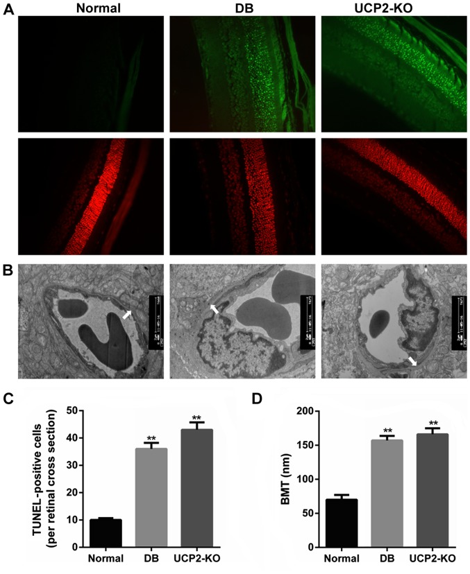
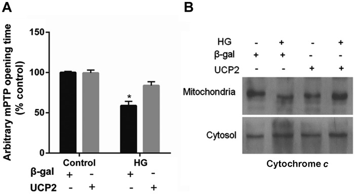
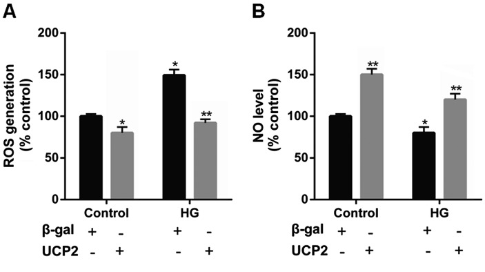
Ectopic apoptosis of vascular cells plays a critical role in the early stage development of diabetic retinopathy (DR). Uncoupling protein 2 (UCP2) is a mitochondrial modulator which protects against endothelial dysfunction. However, the role which UCP2 plays in endothelial apoptosis and its association with DR was unclear. In the present study, we investigated whether UCP2 functioned as an inhibitor of DR in endothelial cells. Firstly, we noted that in UCP2‑knockout mice retinal cell death and damage in vivo was similar to that of db/db diabetic mice. Additionally, UCP2 knockdown induced caspase-3 activation and exaggerated high glucose (HG)-induced apoptosis of human umbilical vein endothelial cells (HUVECs). Conversely, adenovirus-mediated UCP2 overexpression inhibited the apoptosis of HUVECs and HG-induced caspase-3 activation. Furthermore, HG treatment resulted in the opening of the permeability transition pore (PTP) and liberation of cytochrome c from mitochondria to the cytosol in HUVECs. Notably, UCP2 overexpression inhibited these processes. Furthermore, adenovirus-mediated UCP2 overexpression led to a significant increase in intracellular nitric oxide (NO) levels and a decrease in reactive oxygen species (ROS) generation in HUVECs. Collectively, these data suggest that UCP2 plays an anti-apoptotic role in endothelial cells. Thus, we suggest that approaches which augment UCP2 expression in vascular endothelial cells aid in preventing the early stage development and progression of DR.


Figures

Figure 1
Figure 1
Effect of uncoupling protein 2 (UCP2) on retinal cell death and damage in UCP2-knockout (KO) mice. (A) Retinal distribution of TUNEL-positive cells in normal, db/db, and UCP2-KO mice. Sections were counterstained with PI. For clearer TUNEL identification, the retinal area of the diabetic animal was studied using separated and merged TUNEL and PI staining. (B) Transmission electron microscopy of a capillary from the outer plexiform layer of the mouse retina in the three groups. Arrows denote the segment of the outer capillary BM between the endothelial cells and glia limitans, which was used to measure basement membrane width (original magnification, ×9,000). (C) Retinal quantitation of TUNEL-positive cells. TUNEL-positive cells were counted in 10 adjacent locations along the vertical meridian within 4 mm of the optic disk (10 fields/retina section). (D) Retinal capillaries (basement membrane thickness, nm) in the three groups. Data are represented as the means ± SD from eight mice per group, and the experiments were repeated independently at least three times with similar results. Normal, C57 mice. **P<0.01 vs. control.
Figure 2
Figure 2
Effect of uncoupling protein 2 (UCP2)-knockdown on high glucose (HG)-induced apoptosis and caspase-3 activation in human umbilical vein endothelial cells (HUVECs). (A) Two days after transfection, UCP2 protein expression was examined by western blot analysis. (B) Transfected cells were maintained in media containing 0.5% (v/v) FBS for 12 h, and 30 M HG for 16 h. Following treatment, caspase-3 activation and apoptosis were analyzed using a caspase fluorescence assay kit. (C) Transfected cells treated with HG were double stained with Annexin V and PI using the Annexin V-FITC apoptosis detection kit I. The proportion of apoptotic and necrotic cells was determined by FACSCalibur. (D) Effect of UCP overexpression on HG-induced apoptosis and necrosis. Apoptotic and necrotic cells were identified by staining with Annexin V (early apoptosis) or Annexin V/PI (late apoptosis) and PI, respectively. *P<0.01 vs. control-siRNA and **P<0.01 vs. Control-UCP2-siRNA.
Figure 3
Figure 3
Subcellular localization of uncoupling protein 2 (UCP2) protein in UCP2-overexpressing human umbilical vein endothelial cells (HUVECs). (A) Overexpression of UCP2 in HUVECs by transient infection with recombinant adenovirus constructs containing UCP2 sense cDNA and eGFP (Ad-UCP2-eGFP). Live cell images were taken under an inverted epifluorescence microscope, showing green fluorescence for overexpressed eGFP-UCP2 for the different concentrations of adenovirus-UCP2-eGFP (MOI of 1, 5 and 10 pfu/cell, respectively). (B) Adenovirus-mediated gene transfer (6×10⁶ pfu/ml) to confluent HUVECs was performed by 1-h infection at 37°C in ECM without serum. After 48 h, western blot analysis was performed on cytosolic and mitochondrial preparations using an antibody against human UCP2 and subunit IV of cytochrome oxidase, a standard mitochondrial marker. UCP2 was overexpressed mainly within mitochondria. (C) UCP2 and COX IV protein levels were analyzed. Cyt/cyto, cytosol; mt, mitochondria. The values are presented as the means ± SD of three independent experiments. **P<0.01 vs. mt+β-gal.
Figure 4
Figure 4
Effect of uncoupling protein 2 (UCP2) overexpression on high glucose (HG)-induced apoptosis and caspase activation in human umbilical vein endothelial cells (HUVECs). (A) Ad-UCP2 or adenovirus β-galactosidase (Ad-β-gal)-infected HUVECs treated with HG were double stained with Annexin V and PI using the Annexin V-FITC apoptosis detection kit. The proportion of apoptotic and necrotic cells was determined by FACSCalibur. (B) Effect of UCP2 overexpression on HG-induced apoptosis and necrosis. Apoptotic and necrotic cells were identified by staining with Annexin V (early apoptosis) or Annexin V/PI (late apoptosis) and PI, respectively. (C) Western blot analysis for the active subunit of caspase-3. (D) Caspase-3 activation was analyzed by measuring the activity using a caspase-3 colorimetric protease assay kit. The values are presented as the means ± SD of three independent experiments. *P<0.01 vs. control and **P<0.01 vs. β-gal.
Figure 5
Figure 5
Effect of uncoupling protein 2 (UCP2) overexpression on per meability transition pore (PTP) opening and cytochrome c release. (A) Overexpression of UCP2 in human umbilical vein endothelial cells (HUVECs) significantly delayed the time of mPTP opening compared to the adenovirus β-galactosidase (Ad-β-gal) control. (B) Western blot analysis of the release of cytochrome c from mitochondria into the cytosol. The values are presented as the means ± SD of three independent experiments. *P<0.01 vs. control.
Figure 6
Figure 6
Effect of uncoupling protein 2 (UCP2) overexpression on reactive oxygen species (ROS) generation and NO production in human umbilical vein endothelial cells (HUVECs). Quiescent HUVECs were infected with Ad-UCP2 or Ad-β-gal. Two days after infection, cells were maintained in media containing 0.5% (v/v) FBS for 12 h, and exposed to 30 M high glucose (HG) for 6 h. (A) After 15 min incubation with 10 µmol/ml DCFH2-DA, the increase in DCFH2 oxidation was measured using FACSCalibur. (B) NO levels in HUVECs were measured in situ using DAF-FM diacetate. Values are presented as the means ± SD of three independent experiments. *P<0.01 vs. control and **P<0.01 vs. β-gal.

References

    1. Cerani A, Tetreault N, Menard C, Lapalme E, Patel C, Sitaras N, Beaun F, Leboeuf D, De Guire V, Binet F, et al. Neuron-derived semaphorin 3A is an early inducer of vascular permeability in diabetic retinopathy via neuropilin-1. Cell Metab. 2013;18:505–518. doi: 10.1016/j.cmet.2013.09.003. - DOI - PubMed
    1. Barot M, Gokulgandhi MR, Patel S, Mitra AK. Microvascular complications and diabetic retinopathy: recent advances and future implications. Future Med Chem. 2013;5:301–314. doi: 10.4155/fmc.12.206. - DOI - PMC - PubMed
    1. Geraldes P, Hiraoka-Yamamoto J, Matsumoto M, Clermont A, Leitges M, Marette A, Aiello LP, Kern TS, King GL. Activation of PKC-delta and SHP-1 by hyperglycemia causes vascular cell apoptosis and diabetic retinopathy. Nat Med. 2009;15:1298–1306. doi: 10.1038/nm.2052. - DOI - PMC - PubMed
    1. Jiang Y, Zhang Q, Soderland C, Steinle JJ. TNFα and SOCS3 regulate IRS-1 to increase retinal endothelial cell apoptosis. Cell Signal. 2012;24:1086–1092. doi: 10.1016/j.cellsig.2012.01.003. - DOI - PMC - PubMed
    1. Montero JA, Ruiz-Moreno JM, Correa ME. Intravitreal anti-VEGF drugs as adjuvant therapy in diabetic retinopathy surgery. Curr Diabetes Rev. 2011;7:176–184. doi: 10.2174/157339911795843104. - DOI - PubMed

Publication types