

Consecutive bilateral decompression retinopathy after mitomycin C trabeculectomy: a case report
- PMID: 26846338
- PMCID: PMC4743165
- DOI: 10.1186/s13256-016-0814-x
Consecutive bilateral decompression retinopathy after mitomycin C trabeculectomy: a case report
Abstract
Background: After a successful trabeculectomy, a sudden intraocular pressure decrease may alter the intracranial to intraocular pressure ratio and cause decompression retinopathy. Frequent Valsalva maneuvers may also play a role in its pathogenesis. This condition may manifest as multiple retinal hemorrhages, edema of the optic disc, macular edema, or a sudden decrease in visual acuity postoperatively. Outcomes for patients are usually good, with spontaneous resolution occurring within a matter of weeks. It has been rarely reported in the literature as a bilateral condition.
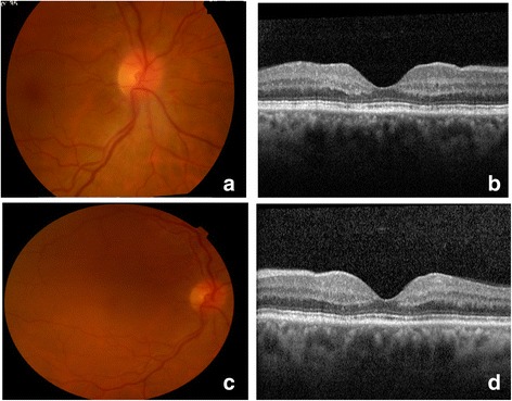
Case presentation: We present a case of consecutive bilateral decompression retinopathy in a 54-year-old severely obese Caucasian woman (body mass index 37 kg/m(2)) with open angle glaucoma and a poor history of medical therapeutic compliance, who chose surgical treatment based on her inability to consistently use ocular drops. Our patient underwent a trabeculectomy with mitomycin C in both eyes, with surgeries taking place 3 months apart. After the first surgery, 2 weeks postoperatively, she complained of decreased visual acuity. Examination of her right eye fundus revealed multiple retinal hemorrhages and disc edema. There was a similar pattern in her left eye, this time including maculopathy. Her visual acuity and fundoscopic changes resolved spontaneously over a period of a month in both cases. Currently, our patient has well-controlled bilateral intraocular pressure, ranging between 14 and 16 mmHg, without hypotensive medication.
Conclusions: Decompression retinopathy is a potential complication after glaucoma surgery, but has rarely been described as a bilateral consecutive condition. A comprehensive approach could help to anticipate its occurrence and manage it.
Figures


Similar articles
-
[Decompression retinopathy with maculopathy after trabeculectomy with mitomycin C].Arch Soc Esp Oftalmol. 2008 Jun;83(6):373-6. doi: 10.4321/s0365-66912008000600007. Arch Soc Esp Oftalmol. 2008. PMID: 18521770 Spanish.
-
Decompression Retinopathy Following Trabeculectomy: A Case Report.Nepal J Ophthalmol. 2020 Jul;12(24):323-327. doi: 10.3126/nepjoph.v12i2.29247. Nepal J Ophthalmol. 2020. PMID: 33978628
-
Decompression retinochoroidopathy: a unique presentation post-trabeculectomy.Int Ophthalmol. 2019 Apr;39(4):927-928. doi: 10.1007/s10792-018-0871-9. Epub 2018 Mar 10. Int Ophthalmol. 2019. PMID: 29525904
-
Ocular decompression retinopathy after deep sclerectomy with mitomycin C in an eye with exfoliation glaucoma.Eur J Ophthalmol. 2011 May-Jun;21(3):324-7. doi: 10.5301/EJO.2010.5731. Eur J Ophthalmol. 2011. PMID: 20872360
-
Three types of retinal bleeding as a complication of hypotony after trabeculectomy.Ophthalmologica. 1999;213(2):135-8. doi: 10.1159/000027407. Ophthalmologica. 1999. PMID: 9885392 Review.
Cited by
-
Bilateral Ocular Decompression Retinopathy after Ahmed Valve Implantation for Uveitic Glaucoma.Case Rep Ophthalmol. 2016 Nov 1;7(3):227-232. doi: 10.1159/000452268. eCollection 2016 Sep-Dec. Case Rep Ophthalmol. 2016. PMID: 27920718 Free PMC article.
References
Publication types
MeSH terms
Substances
LinkOut - more resources
Full Text Sources
Other Literature Sources
Medical
