Review
. 2016 Apr;173(2):206-18.
doi: 10.1111/bjh.13938. Epub 2016 Feb 5.

Advances in understanding erythropoiesis: evolving perspectives

Affiliations
Review

Advances in understanding erythropoiesis: evolving perspectives

Satish K Nandakumar et al. Br J Haematol. 2016 Apr.

Abstract

Red blood cells (RBCs) are generated from haematopoietic stem and progenitor cells (HSPCs) through the step-wise process of differentiation known as erythropoiesis. In this review, we discuss our current understanding of erythropoiesis and highlight recent advances in this field. During embryonic development, erythropoiesis occurs in three distinct waves comprising first, the yolk sac-derived primitive RBCs, followed sequentially by the erythro-myeloid progenitor (EMP) and HSPC-derived definitive RBCs. Recent work has highlighted the complexity and variability that may exist in the hierarchical arrangement of progenitors responsible for erythropoiesis. Using recently defined cell surface markers, it is now possible to enrich for erythroid progenitors and precursors to a much greater extent than has been possible before. While a great deal of knowledge has been gained on erythropoiesis from model organisms, our understanding of this process is currently being refined through human genetic studies. Genes mutated in erythroid disorders can now be identified more rapidly by the use of next-generation sequencing techniques. Genome-wide association studies on erythroid traits in healthy populations have also revealed new modulators of erythropoiesis. All of these recent developments have significant promise not only for increasing our understanding of erythropoiesis, but also for improving our ability to intervene when RBC production is perturbed in disease.

Keywords: erythropoiesis; haematopoiesis; haemopoietic progenitors; red cell disorders; red cells.


Figures

Figure 1
Figure 1
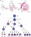
A) There are three developmental waves of erythropoiesis in mammals. The first wave is marked by the emergence of primitive erythroblasts (PE) that express embryonic globins in yolk sac blood islands. In the second wave, the erythro-myeloid progenitor (EMP) emerges from yolk sac and migrates to the fetal liver, producing definitive erythroblasts expressing predominantly mouse adult globins. In the third wave, the haematopoietic stem cell (HSC) emerges from the haemogenic endothelium in the aorto-gonad mesonephros (AGM) and other sites. The HSC migrates to fetal liver and eventually to the adult bone marrow (BM), producing definitive erythroblasts. B) Classical (in black) and alternate (in red) models of the adult haematopoietic hierarchy. In the classical model, the adult HSCs in the BM gives rise to either a common myeloid progenitor (CMP) or common lymphoid progenitor (CLP). The CMP then differentiates into either a granulocyte monocyte progenitor (GMP) or megakaryocyte erythroid progenitor (MEP). These progenitors differentiate into mature cells of distinct lineages. Several alternate pathways have been discovered by recent studies (in red). HSCs were shown to differentiate directly into CMP, MEP and megakaryocytes. HSC can also differentiate into a lymphoid primed multipotent progenitor (LMPP) lacking any megakaryocyte erythroid potential. NK, Natural killer cell; LT-HSC, long-term HSC; ST-HSC; short-term HSC; MPP, multipotent progenitor; RBC, red blood cell.
Figure 2
Figure 2
The differentiation steps from the megakaryocyte erythroid progenitor (MEP) to the mature red blood cell (RBC) are shown. An overview of recent strategies to isolate these cell types based on surface markers is given. Note that the current MEP isolation strategies also appear to contain BFU-Es and CFU-Es. MEP, Megakaryocyte erythroid progenitor; BFU-E, blast colony forming unit - erythroid; CFU-E, colony forming unit - erythroid; ProE, proerythroblast; BasoE, basophilic erythroblast; PolyE, polychromatic erythroblast; OrthoE, orthochromatic erythroblast; Retic, reticulocyte.
Figure 3
Figure 3
Brief overview of the advantages and disadvantages of model systems commonly used to understand erythropoiesis. PSC, pluripotent stem cell.
Figure 4
Figure 4
A) The core erythroid network (CEN) of transcription factors (TFs) is composed of the DNA-binding TFs, GATA1, TAL1 and KLF1, as well as the non-DNA-binding TFs, LDB1 and LMO2. The CEN loops from enhancers to promoters to activate target gene expression. B) TFs in the CEN often interact with additional TFs at a subset of regulatory elements. For example, GATA1 and TAL1 interact with GFI1B and the nucleosome remodelling deacetylase (NuRD) complex, resulting in target gene repression. C) Long non-coding RNAs (lncRNAs) that are transcribed during erythropoiesis are regulated by the CEN. Future studies will probably refine our understanding of the role of lncRNAs in erythropoiesis.

References

    1. Alvarez-Dominguez JR, Hu W, Yuan B, Shi J, Park SS, Gromatzky AA, van Oudenaarden A, Lodish HF. Global discovery of erythroid long noncoding RNAs reveals novel regulators of red cell maturation. Blood. 2014;123:570–581. - PMC - PubMed
    1. Arlet JB, Ribeil JA, Guillem F, Negre O, Hazoume A, Marcion G, Beuzard Y, Dussiot M, Moura IC, Demarest S, de Beauchene IC, Belaid-Choucair Z, Sevin M, Maciel TT, Auclair C, Leboulch P, Chretien S, Tchertanov L, Baudin-Creuza V, Seigneuric R, Fontenay M, Garrido C, Hermine O, Courtois G. HSP70 sequestration by free alpha-globin promotes ineffective erythropoiesis in beta-thalassaemia. Nature. 2014;514:242–246. - PubMed
    1. Basak A, Hancarova M, Ulirsch JC, Balci TB, Trkova M, Pelisek M, Vlckova M, Muzikova K, Cermak J, Trka J, Dyment DA, Orkin SH, Daly MJ, Sedlacek Z, Sankaran VG. BCL11A deletions result in fetal hemoglobin persistence and neurodevelopmental alterations. Journal of Clinical Investigation. 2015;125:2363–2368. - PMC - PubMed
    1. Bassett AR, Akhtar A, Barlow DP, Bird AP, Brockdorff N, Duboule D, Ephrussi A, Ferguson-Smith AC, Gingeras TR, Haerty W, Higgs DR, Miska EA, Ponting CP. Considerations when investigating lncRNA function in vivo. Elife. 2014;3:e03058. - PMC - PubMed
    1. Bauer DE, Kamran SC, Lessard S, Xu J, Fujiwara Y, Lin C, Shao Z, Canver MC, Smith EC, Pinello L, Sabo PJ, Vierstra J, Voit RA, Yuan GC, Porteus MH, Stamatoyannopoulos JA, Lettre G, Orkin SH. An erythroid enhancer of BCL11A subject to genetic variation determines fetal hemoglobin level. Science. 2013;342:253–257. - PMC - PubMed

Publication types