

Evaluation of BP-ONJ in osteopenic and healthy sheep: comparing ZTE-MRI with µCT
- PMID: 26846710
- PMCID: PMC4846170
- DOI: 10.1259/dmfr.20150250
Evaluation of BP-ONJ in osteopenic and healthy sheep: comparing ZTE-MRI with µCT
Abstract
Objectives: Bisphosphonate-associated osteonecrosis of the jaw (BP-ONJ) is a side effect of antiresorptive treatment that is increasingly prescribed for patients with osteoporosis or malignant diseases with bone metastases. Surgical treatment of BP-ONJ requires adequate pre-operative imaging. To date, CT is the imaging standard in clinical routine; however, defining the extent of the pathological area is difficult and soft tissues are poorly displayed. MRI with zero echo time (ZTE-MRI) to display hard tissues enables a precise display of calcified structures and soft tissues for the delineation of bone necrosis and soft-tissue reactions.
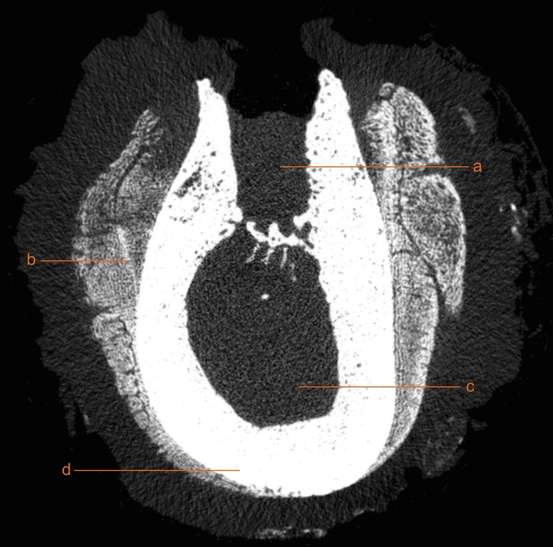
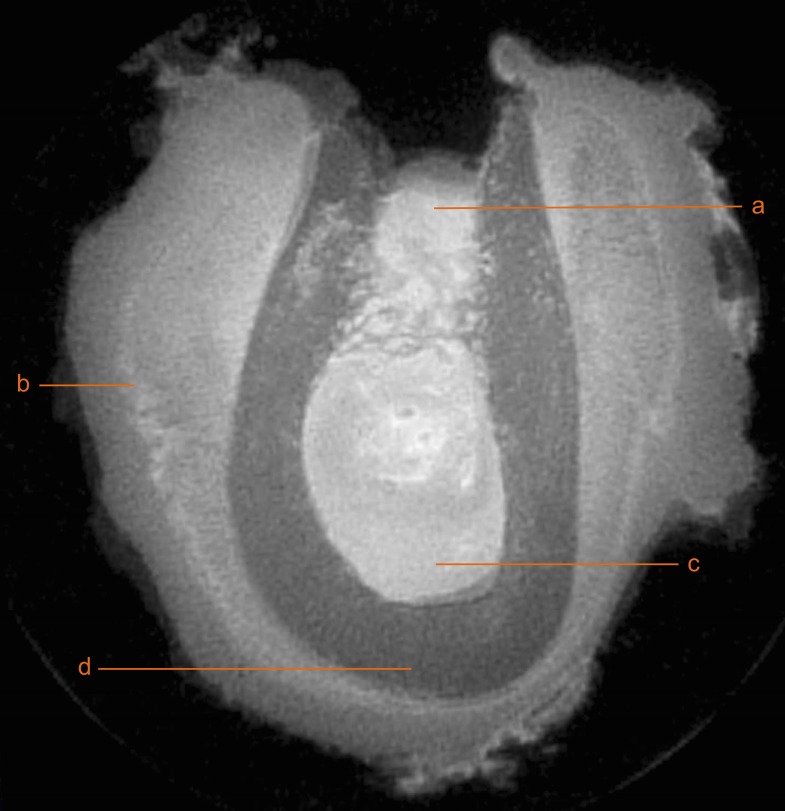
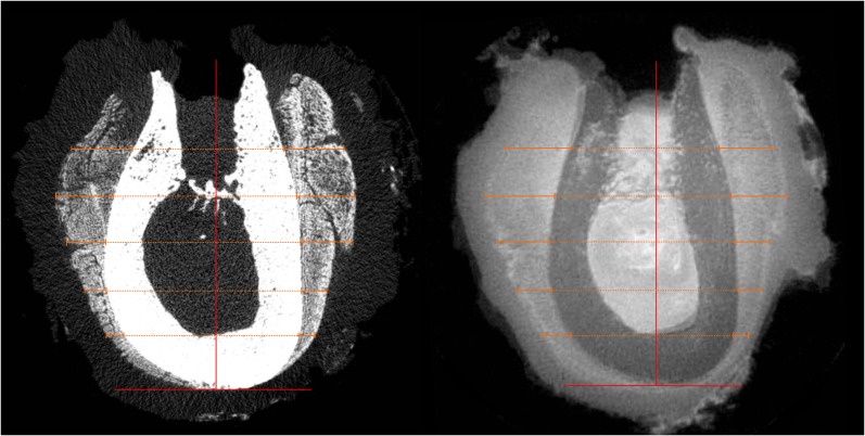
Methods: BP-ONJ was induced in eight sheep by extraction of two premolars in the left mandible and zoledronate (ZOL) administration. Eight sheep without ZOL administration served as the control group. Four sheep of each main group underwent osteopenia induction via ovariectomy, glucocorticoid administration and a calcium-free diet. After sacrifice, the area of tooth extraction was harvested and scanned with micro-CT (µCT) and ZTE-MRI. Two trained dentists analyzed digital imaging and communications in medicine data sets using three-dimensional imaging software. The periosteal reaction and the remaining extraction sockets were measured.
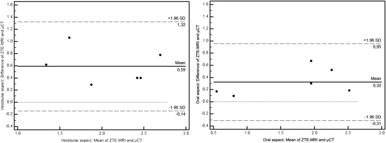
Results: BP-ONJ was evident, and the remaining extraction sockets were observed in all animals treated with ZOL. Periosteal reactions were more pronounced in animals treated with ZOL, and they appeared broader in ZTE-MRI.
Conclusions: BP-ONJ lesions in the sheep mandible can be detected using µCT and ZTE-MRI. Although illustration of sequester was more consistent using the µCT, ZTE-MRI was advantageous in evaluation of periosteal reaction.
Keywords: animal model; bisphosphonate-associated osteonecrosis of the jaw; magnetic resonance imaging; periosteum; x-ray microtomography.
Figures

Similar articles
-
Zoledronate induces bisphosphonate-related osteonecrosis of the jaw in osteopenic sheep.Clin Oral Investig. 2016 Jan;20(1):31-8. doi: 10.1007/s00784-015-1468-6. Epub 2015 Apr 7. Clin Oral Investig. 2016. PMID: 25843053
-
Lansoprazole and zoledronate delays hard tissue healing of tooth extraction sockets in dexamethasone-treated mice.Biomed Pharmacother. 2022 Jun;150:112991. doi: 10.1016/j.biopha.2022.112991. Epub 2022 Apr 21. Biomed Pharmacother. 2022. PMID: 35462336
-
Zoledronate induces osteonecrosis of the jaw in sheep.J Craniomaxillofac Surg. 2015 Sep;43(7):1133-8. doi: 10.1016/j.jcms.2015.04.020. Epub 2015 Apr 30. J Craniomaxillofac Surg. 2015. PMID: 26154396
-
Periodontal disease preceding osteonecrosis of the jaw (ONJ) in cancer patients receiving antiresorptives alone or combined with targeted therapies: report of 5 cases and literature review.Oral Surg Oral Med Oral Pathol Oral Radiol. 2015 Dec;120(6):699-706. doi: 10.1016/j.oooo.2015.08.007. Epub 2015 Aug 21. Oral Surg Oral Med Oral Pathol Oral Radiol. 2015. PMID: 26455289 Review.
-
Osteonecrosis of the Jaw in Patients Receiving Bone-Targeted Therapies: An Overview--Part I.Urol Nurs. 2016 May-Jun;36(3):111-6, 154. Urol Nurs. 2016. PMID: 27501591 Review.
Cited by
-
Pathogenesis of medication-related osteonecrosis of the jaw: a comparative study of in vivo and in vitro trials.J Int Med Res. 2018 Oct;46(10):4277-4296. doi: 10.1177/0300060518788987. Epub 2018 Aug 9. J Int Med Res. 2018. PMID: 30091399 Free PMC article. Review.
-
Microcirculatory consequences of limb ischemia/reperfusion in ovariectomized rats treated with zoledronic acid.J Orthop Surg Res. 2019 Apr 4;14(1):95. doi: 10.1186/s13018-019-1117-x. J Orthop Surg Res. 2019. PMID: 30947735 Free PMC article.
-
A large animal model of periodontal defects in bisphosphonate-related osteonecrosis of the jaw: a comparison of clinical and radiological findings.J Periodontal Implant Sci. 2024 Jun;54(3):139-148. doi: 10.5051/jpis.2204860243. Epub 2023 Jul 7. J Periodontal Implant Sci. 2024. PMID: 37681353 Free PMC article.
References
-
- Ruggiero SL, Dodson TB, Fantasia J, Goodday R, Aghaloo T, Mehrotra B, et al. ; American Association of Oral and Maxillofacial Surgeons. American Association of Oral and Maxillofacial Surgeons position paper on medication-related osteonecrosis of the jaw–2014 update. J Oral Maxillofac Surg 2014; 72: 1938–56. doi: http://dx.doi.org/10.1016/j.joms.2014.04.031 - DOI - PubMed
-
- Patel S, Choyee S, Uyanne J, Nguyen AL, Lee P, Sedghizadeh PP, et al. . Non-exposed bisphosphonate-related osteonecrosis of the jaw: a critical assessment of current definition, staging, and treatment guidelines. Oral Dis 2012; 18: 625–32. doi: http://dx.doi.org/10.1111/j.1601-0825.2012.01911.x - DOI - PubMed
-
- Dore F, Filippi L, Biasotto M, Chiandussi S, Cavalli F, Di Lenarda R. Bone scintigraphy and SPECT/CT of bisphosphonate-induced osteonecrosis of the jaw. J Nucl Med 2009; 50: 30–5. doi: http://dx.doi.org/10.2967/jnumed.107.048785 - DOI - PubMed
-
- Dixon R, Tricker N, Garetto L, eds. Bone turnover in elderly canine mandible and tibia. J Dent Res, ALEXANDRIA, VA: AMER ASSOC DENTAL RESEARCH; 1997.
-
- Van den Wyngaert T, Huizing MT, Fossion E, Vermorken JB. Scintigraphic evaluation of mandibular bone turnover in patients with solid tumors receiving zoledronic acid. Oral Oncol 2010; 46: 214–8. doi: http://dx.doi.org/10.1016/j.oraloncology.2010.01.001 - DOI - PubMed
Publication types
MeSH terms
Substances
LinkOut - more resources
Full Text Sources
Other Literature Sources
Medical
