. 2016 Apr 15;291(16):8516-27.
doi: 10.1074/jbc.M115.707968. Epub 2016 Feb 4.

Preparation and Characterization of Stable α-Synuclein Lipoprotein Particles

Affiliations

Preparation and Characterization of Stable α-Synuclein Lipoprotein Particles

Cédric Eichmann et al. J Biol Chem. .

Abstract

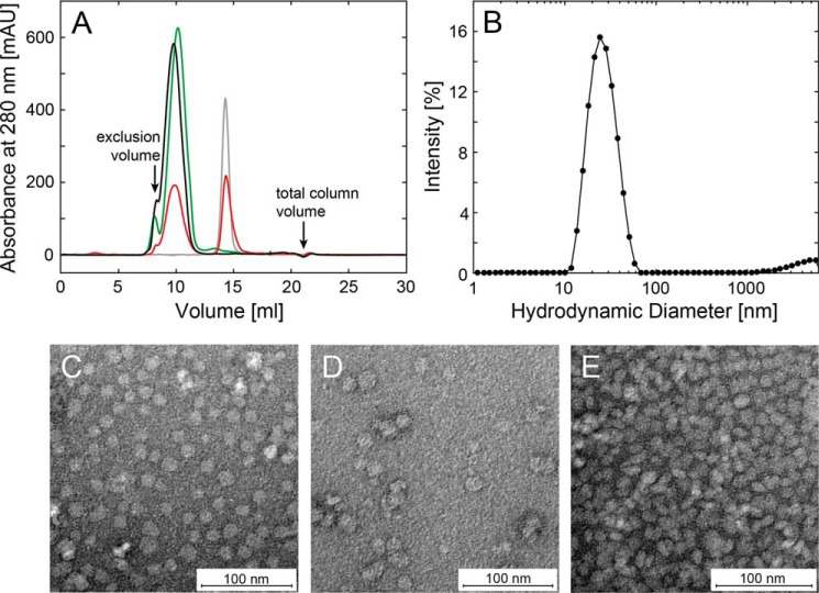
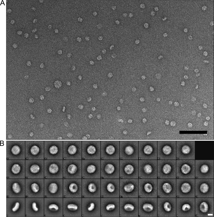
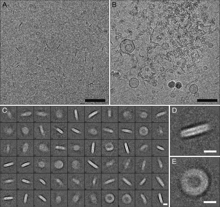
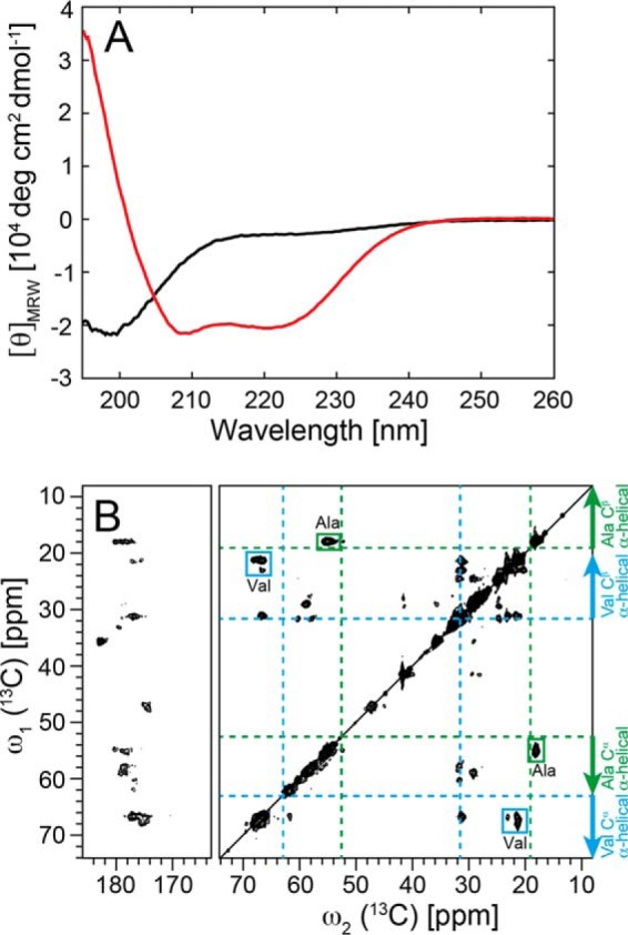
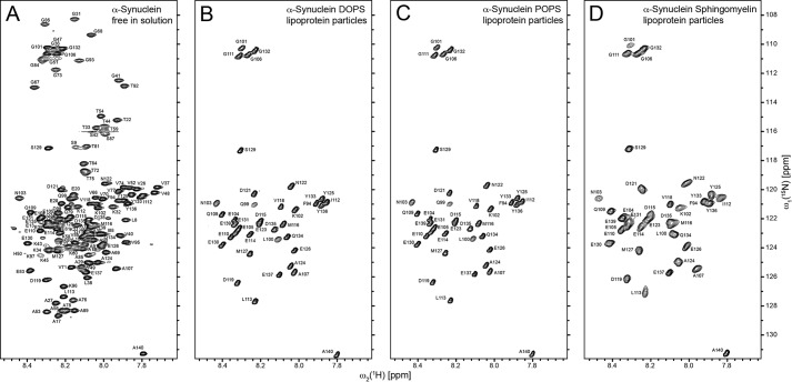
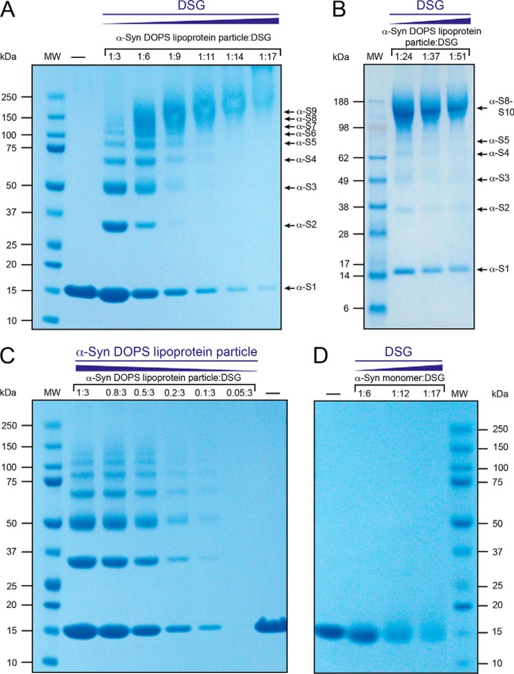
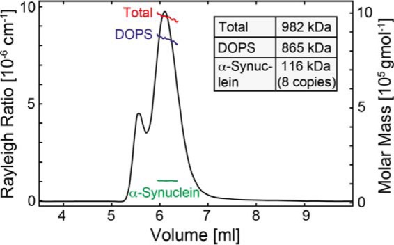
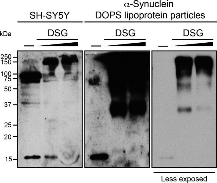
Multiple neurodegenerative diseases are caused by the aggregation of the human α-Synuclein (α-Syn) protein. α-Syn possesses high structural plasticity and the capability of interacting with membranes. Both features are not only essential for its physiological function but also play a role in the aggregation process. Recently it has been proposed that α-Syn is able to form lipid-protein particles reminiscent of high-density lipoproteins. Here, we present a method to obtain a stable and homogeneous population of nanometer-sized particles composed of α-Syn and anionic phospholipids. These particles are called α-Syn lipoprotein (nano)particles to indicate their relationship to high-density lipoproteins formed by human apolipoproteins in vivo and of in vitro self-assembling phospholipid bilayer nanodiscs. Structural investigations of the α-Syn lipoprotein particles by circular dichroism (CD) and magic angle solid-state nuclear magnetic resonance (MAS SS-NMR) spectroscopy establish that α-Syn adopts a helical secondary structure within these particles. Based on cryo-electron microscopy (cryo-EM) and dynamic light scattering (DLS) α-Syn lipoprotein particles have a defined size with a diameter of ∼23 nm. Chemical cross-linking in combination with solution-state NMR and multiangle static light scattering (MALS) of α-Syn particles reveal a high-order protein-lipid entity composed of ∼8-10 α-Syn molecules. The close resemblance in size between cross-linked in vitro-derived α-Syn lipoprotein particles and a cross-linked species of endogenous α-Syn from SH-SY5Y human neuroblastoma cells indicates a potential functional relevance of α-Syn lipoprotein nanoparticles.

Keywords: Parkinson disease; alpha-synuclein (α-synuclein); apolipoprotein; high-density lipoprotein (HDL); lipid; membrane bilayer.


Figures

FIGURE 1.
FIGURE 1.
α-Syn-containing lipoprotein particles are well defined in size. A, size-exclusion gel chromatography (Superdex 200 10/300GL) of α-Syn DOPS (black), α-Syn POPS (green), and α-Syn sphingomyelin (red) lipoprotein particles. Monomeric (gray) α-Syn elutes at ∼14.3 ml. B, dynamic light scattering of α-Syn-containing DOPS lipoprotein particles. The size-exclusion chromatography fraction at the maximum of the absorbance of 280 nm (9.5–10 ml) consists of particles with an average hydrodynamic radius of 25.8 ± 0.8 nm. C–E, negatively stained electron micrographs containing the maxima of the size-exclusion chromatography fractions at the absorbance of 280 nm of purified (C) α-Syn DOPS, (D) α-Syn POPS, and (E) α-Syn sphingomyelin lipoprotein particles. The radius varies from ∼19–28 nm, scale bar 100 nm.
FIGURE 2.
FIGURE 2.
Electron microscopy of α-Syn DOPS lipoprotein particles. A, electron micrograph of negatively stained α-Syn lipoprotein particles. Both top and side views of the nanoparticles are visible (scale bar 100 nm). B, two-dimensional classification of α-Syn lipoprotein particles (scale bar 10 nm).
FIGURE 3.
FIGURE 3.
Cryo-electron microscopy images of α-Syn DOPS lipoprotein particles. A, on the cryo-electron micrograph mostly side views of α-Syn DOPS lipoprotein particles are well visible. B, α-Syn DOPS lipoprotein particles have a tendency to aggregate. C, two-dimensional classification of α-Syn DOPS lipoprotein particles. Three main populations of nanoparticles with diameters of 19–21 nm, 23–24 nm, 27–28 nm, respectively, were observed. D, side and E, top view of two-dimensional nanoparticle classes with a diameter of ∼23 nm. The scale bars indicate 100 nm in (A, B) and 10 nm in (C–E).
FIGURE 4.
FIGURE 4.
Helical secondary structure of α-Syn in lipoprotein particles. A, conformational change of α-Syn determined by CD from a predominantly unfolded state (black) free in solution to an α-helical state in α-Syn DOPS lipoprotein particles (red). For the α-Syn DOPS lipoprotein particles, the size exclusion chromatography fraction 9.5–10 ml (Fig. 1A) was used. B, two-dimensional 13C DARR solid-state NMR spectra (at a magnetic field of 850 MHz 1H frequency) of uniformly 13C, 15N-labeled α-Syn DOPS lipoprotein particles. The assigned cross peaks from Ala and Val residues are indicated by green and cyan boxes, respectively. Indicated with dotted green and cyan lines are the random coil 13Cα, 13Cβ chemical shifts for the amino acid residues Ala and Val, respectively. Arrows show the chemical shift area typically observed for α-helical secondary structure.
FIGURE 5.
FIGURE 5.
The C-terminal ∼40 residues of α-Syn are flexible in α-Syn lipoprotein particles as evidenced by two-dimensional [15N,1H]-TROSY spectra. The spectra of (A) 13C, 15N-labeled α-Syn free in solution and 2H, 15N-labeled (B) α-Syn DOPS, (C) α-Syn POPS, and (D) α-Syn sphingomyelin lipoprotein particles in a buffer containing 20 mm Bis-Tris-HCl, pH 7, 20 mm NaCl were measured at a magnetic field of 700 MHz 1H frequency at 30 °C. The cross peaks are labeled with the one-letter amino acid residue code. The slight peak doubling observed is attributed to 2H isotope effects.
FIGURE 6.
FIGURE 6.
α-Syn DOPS lipoprotein particles comprise 8–10 copies of α-Syn as evidenced by cross-linking studies. SDS-PAGE, 4–12% NuPAGE Bis-Tris gel (Invitrogen), of cross-linked α-Syn DOPS lipoprotein particles. A, lane 1, molecular weight marker (MW, Precision Plus ProteinTM Dual Color Standard, BIORAD). Lane 2, (−) Non-cross-linked α-Syn DOPS lipoprotein particles (control). Lanes 3–8, α-Syn DOPS lipoprotein particles (final concentration 83 μm) were exposed to increasing concentrations (molar ratios) of the amine-reactive cross-linker DSG. Arrowheads indicate presumed α-Syn monomer (α-S1) and oligomers (such as α-S2 for dimeric α-Syn). B, lane 1, molecular weight marker (MW, SeeBlue plus2 prestained Standard, Invitrogen). Lanes 2–4, cross-linked α-Syn DOPS lipoprotein particles (final concentration 83 μm) with increasing concentrations of DSG as indicated. Presumed α-Syn monomer and oligomers are indicated by arrowheads. C, lane 1, MW, Precision Plus ProteinTM Dual Color Standard, BIORAD). Lanes 2–7, decreasing concentrations of α-Syn DOPS lipoprotein particles were exposed to a constant concentration of DSG (final concentration 249 μm). Lane 8, (−) non cross-linked α-Syn DOPS lipoprotein particles for control. D, approximately 9 μg monomeric α-Syn was loaded per well. Lane 1, (−) non cross-linked monomeric α-Syn control. Lanes 2–4, cross-linked monomeric α-Syn (final concentration 83 μm) with increasing concentrations of DSG. Lane 5, MW (Precision Plus ProteinTM Dual Color Standard, Biorad).
FIGURE 7.
FIGURE 7.
Analysis of the α-Syn and DOPS composition in α-Syn DOPS lipoprotein particles. Molecular weight analysis of the α-Syn DOPS lipoprotein complex performed by multiangle static light scattering coupled with size-exclusion gel chromatography and refractive index measurements. The black line corresponds to the static light scattering signal at 454 nm of DABMI-labeled α-Syn(C141) in the presence of DOPS lipids; red, blue, and green lines show average molar masses of the complex, the lipid component, and the protein component in the lipoprotein particle, respectively. Following these investigations, the protein mass is ∼116 kDa indicating that α-Syn is of octameric nature in DOPS lipoprotein particles.
FIGURE 8.
FIGURE 8.
In vivo cross-linked endogenous α-Syn in SH-SY5Y human neuroblastoma cells shows a major high molecular weight species of similar size as cross-linked α-Syn DOPS lipoprotein particles. Left panel, SH-SY5Y cells were exposed to increasing concentrations of the amine-reactive cross-linker DSG: (−) non cross-linked as control (lane 1), 1 and 2 mm DSG cross-linked SH-SY5Y cells (lanes 2 and 3). Middle and right panel, (−) non cross-linked (lane 1), 1 and 2 mm DSG cross-linked α-Syn DOPS lipoprotein particles (lanes 2 and 3).

References

    1. Ulusoy A., and Di Monte D. A. (2013) α-Synuclein elevation in human neurodegenerative diseases: Experimental, pathogenetic, and therapeutic implications. Mol. Neurobiol. 47, 484–494 - PubMed
    1. Spillantini M. G., and Goedert M. (2000) The α-Synucleinopathies: Parkinson's Disease, Dementia with Lewy Bodies, and Multiple System Atrophy. Ann. N.Y. Acad. Sci. 920, 16–27 - PubMed
    1. Weinreb P. H., Zhen W., Poon A. W., Conway K. A., and Lansbury P. T. (1996) NACP, a protein implicated in Alzheimer's disease and learning, is natively unfolded. Biochemistry 35, 13709–13715 - PubMed
    1. Uversky V. N. (2003) A protein-chameleon: conformational plasticity of α-synuclein, a disordered protein involved in neurodegenerative disorders. J. Biomol. Struct. Dyn. 21, 211–234 - PubMed
    1. Eliezer D., Kutluay E., Bussell R. Jr., and Browne G. (2001) Conformational properties of α-synuclein in its free and lipid-associated states. J. Mol. Biol. 307, 1061–1073 - PubMed

Publication types

LinkOut - more resources