

Predicting Intracerebral Hemorrhage Growth With the Spot Sign: The Effect of Onset-to-Scan Time
- PMID: 26846857
- PMCID: PMC4766058
- DOI: 10.1161/STROKEAHA.115.012012
Predicting Intracerebral Hemorrhage Growth With the Spot Sign: The Effect of Onset-to-Scan Time
Abstract
Background and purpose: Hematoma expansion after acute intracerebral hemorrhage is common and is associated with early deterioration and poor clinical outcome. The computed tomographic angiography (CTA) spot sign is a promising predictor of expansion; however, frequency and predictive values are variable across studies, possibly because of differences in onset-to-CTA time. We performed a patient-level meta-analysis to define the relationship between onset-to-CTA time and frequency and predictive ability of the spot sign.
Methods: We completed a systematic review for studies of CTA spot sign and hematoma expansion. We subsequently pooled patient-level data on the frequency and predictive values for significant hematoma expansion according to 5 predefined categorized onset-to-CTA times. We calculated spot-sign frequency both as raw and frequency-adjusted rates.
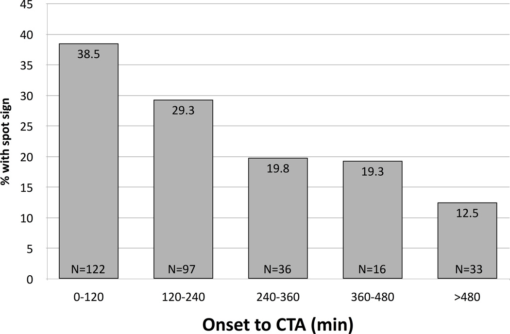
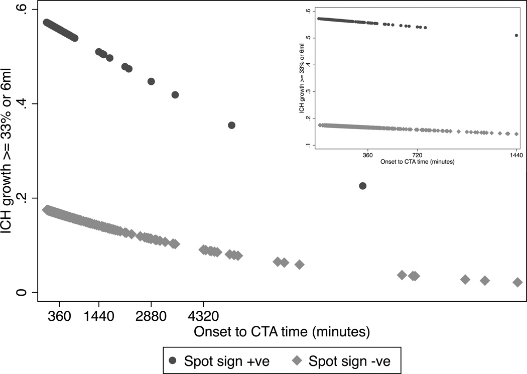
Results: Among 2051 studies identified, 12 met our inclusion criteria. Baseline hematoma volume, spot-sign status, and time-to-CTA were available for 1176 patients, and 1039 patients had follow-up computed tomographies for hematoma expansion analysis. The overall spot sign frequency was 26%, decreasing from 39% within 2 hours of onset to 13% beyond 8 hours (P<0.001). There was a significant decrease in hematoma expansion in spot-positive patients as onset-to-CTA time increased (P=0.004), with positive predictive values decreasing from 53% to 33%.
Conclusions: The frequency of the CTA spot sign is inversely related to intracerebral hemorrhage onset-to-CTA time. Furthermore, the positive predictive value of the spot sign for significant hematoma expansion decreases as time-to-CTA increases. Our results offer more precise risk stratification for patients with acute intracerebral hemorrhage and will help refine clinical prediction rules for intracerebral hemorrhage expansion.
Keywords: CT angiography; cerebral hemorrhage; hematoma expansion; intracerebral hemorrhage; spot sign.
© 2016 American Heart Association, Inc.
Figures
References
-
- van Asch CJ, Luitse MJ, Rinkel GJ, van der Tweel I, Algra A, Klijn CJ. Incidence, case fatality, and functional outcome of intracerebral haemorrhage over time, according to age, sex, and ethnic origin: a systematic review and meta-analysis. Lancet Neurol. 2010;9:167–176. - PubMed
-
- Broderick JP, Brott TG, Duldner JE, Tomsick T, Huster G. Volume of intracerebral hemorrhage. A powerful and easy-to-use predictor of 30-day mortality. Stroke. 1993;24:987–993. - PubMed
-
- Hemphill JC, Bonovich DC, Besmertis L, Manley GT, Johnston SC. The ICH score: a simple, reliable grading scale for intracerebral hemorrhage. Stroke. 2001;32:891–897. - PubMed
-
- Leira R, Dávalos A, Silva Y, Gil-Peralta A, Tejada J, Garcia M, et al. Early neurologic deterioration in intracerebral hemorrhage: predictors and associated factors. Neurology. 2004;63:461–467. - PubMed
Publication types
MeSH terms
Grants and funding
LinkOut - more resources
Full Text Sources
Other Literature Sources
Medical
Miscellaneous
