Review
. 2016 Mar;4(2):189-212.
doi: 10.1111/andr.12165. Epub 2016 Feb 4.

The Sertoli cell: one hundred fifty years of beauty and plasticity

Affiliations
Review

The Sertoli cell: one hundred fifty years of beauty and plasticity

L R França et al. Andrology. 2016 Mar.

Abstract

It has been one and a half centuries since Enrico Sertoli published the seminal discovery of the testicular 'nurse cell', not only a key cell in the testis, but indeed one of the most amazing cells in the vertebrate body. In this review, we begin by examining the three phases of morphological research that have occurred in the study of Sertoli cells, because microscopic anatomy was essentially the only scientific discipline available for about the first 75 years after the discovery. Biochemistry and molecular biology then changed all of biological sciences, including our understanding of the functions of Sertoli cells. Immunology and stem cell biology were not even topics of science in 1865, but they have now become major issues in our appreciation of Sertoli cell's role in spermatogenesis. We end with the universal importance and plasticity of function by comparing Sertoli cells in fish, amphibians, and mammals. In these various classes of vertebrates, Sertoli cells have quite different modes of proliferation and epithelial maintenance, cystic vs. tubular formation, yet accomplish essentially the same function but in strikingly different ways.

Keywords: Sertoli cell; amniotes; anamniotes; blood-testis barrier; germ cell; immune privilege; immune tolerance; spermatogenesis; stem cell niche; testis.


Conflict of interest statement

CONFLICT OF INTEREST

The authors have no conflicts of interest to declare.

Figures

Figure 1
Figure 1
Photo of Professor Enrico Sertoli, published in honor of his retirement after 36 years of teaching and research (Negrini et al., 1908) and drawings (Sertoli, 1865) from Sertoli’s original publication (Fig. 1A–D).
Figure 2
Figure 2
Three phases observed in morphological studies of the Sertoli cell. (A) Light microscopy (LM). The image is from mouse seminiferous epithelium, Stage IV (Periodic acid-Schiff’s stain). The Sertoli cell nucleus is euchromatic with a large nucleolus and a single satellite chromocenter. An intimate association of germ cells with the Sertoli cell is displayed with pachytene spermatocytes adjacent to the Sertoli cell cytoplasm and heads of elongated spermatids that are pulled deep into the Sertoli cell crypts and lying next to the apical region of its nucleus. Round spermatids are found near the lumen. Bar = 12 lm. (B) Transmission electron microscopy (TEM). The tissue is from human testis, showing the Sertoli cell resting on the basement membrane and surrounded by germ cells. Spermatogonia (Spg); Spermatocyte (Spc). The Sertoli cell has a highly euchromatic nucleus (SCN), a large nucleolus (Nu) with two satellite chromocenters (Cc) and an indentation of the nuclear membrane (In). Plasmalemma (Pl); Ectoplasmic specialization (Eps) at the blood–testis barrier. Bar = 5 μm. (C) Immunofluorescence microscopy. The Sertoli cell was isolated in vitro with attached elongated spermatids and labeled for somatic cell-specific tubulin (green) and filamentous actin (red). Actin is labeled at the ectoplasmic specialization and the intercellular bridges. The microtubules are continuous with the apical regions and around the basal area of the nucleus and directly adjacent to the ectoplasmic specializations, attached to the spermatid heads that are drawn deep into the Sertoli cell crypts. Bar = 10 μm. Original illustration provided by Dr. A. Wayne Vogl, Department of Cellular and Physiological Sciences, Faculty of Medicine, University of British Columbia, Vancouver, Canada (Vogl et al., 1995). Modified and reprinted with permission of the Copyright © holder Elsevier.
Figure 3
Figure 3
Schematic illustration of the Sertoli cell’s interaction with germ cells at different stages during spermatogenesis and other key functions, including: (1) transport of micronutrients across the junctional complex; (2) management of waste and recycled leftover cytoplasm during germ cell development; (3) maintenance of the blood–testis barrier (BTB); (4) establishment of germ cell adhesions and communication; (5) inhibition of immune reactions and maintenance of immune privilege; (6) initiation and response to endocrine signaling pathways; (7) initiation and regulation of the cycle of the seminiferous epithelium; and (8) maintenance of stem cell homeostasis. Most autoimmunogenic germ cells are sequestered within the adluminal compartment of the seminiferous epithelium behind the BTB, where Sertoli cells surround them. Sertoli cells secrete immunoregulatory factors (5) that modify the immune response and induce regulatory immune cells such as macrophages (M2) and T cells (Tregs). (A) Actin filaments (green) are seen along the basal Sertoli/Sertoli tight junctions but also lining the heads of elongated spermatids; (B) Claudin-11 (red) stains only the basal junctional complex; (C) Actin (green), Rab5 (red) and DAPI (blue for nucleus) show the intricate relationship of these proteins to the tubulobulbar complex; (D) Androgen receptor (brown) stains only the Sertoli cell nucleus in the hamster seminiferous epithelium. Original illustration provided by Dr. Wayne Vogl (Hess & Vogl, 2015). Modified with approval of the Copyright © holder for Sertoli Cell Biology, 2nd edition, Elsevier Academic Press.
Figure 4
Figure 4
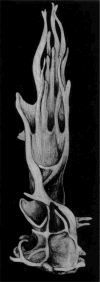
A three-dimensional drawing of a Stage V rat Sertoli cell taken from a photograph of a plastic model created from 675 micrographs of 372 serial electron microscopic sections. Cellular processes and cup-like hollows show the intimate relationship with adjacent germ cells (Wong & Russell, 1983). Reprinted with approval of the Copyright © holder John Wiley & Sons, Inc.
Figure 5
Figure 5
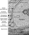
Electron microscopy of adjacent Sertoli cells showing the tight junctional complex (Sertoli–Sertoli tight junction) and associated basal ectoplasmic specialization. The Sertoli cell plasmalemma is seen between the two cells, which sit on the basal lamina and a peritubular myoid cell. One nucleolar chromocenters is noted in the large, euchromatic nucleus.
Figure 6
Figure 6
Electron microscopy showing the thin arm of a Sertoli cell (light green area) containing the ectoplasmic specialization (Eps) and Sertoli cell cytoplasm (Sc) adjacent to the germ cell cytoplasm showing the manchette microtubules of spermatid 2 and the acrosome (Ac) that covers the nucleus (N) of spermatid 1.
Figure 7
Figure 7
Model depicting the possible role of NOTCH signaling in Sertoli cells after birth. Previous studies have shown that FGF2 and FSH induce GDNF expression by Sertoli cells. Recent data suggest NOTCH signaling is a negative regulator of GDNF, which might balance the effects of FGF2 and FSH. Overactivation of NOTCH signaling suppresses the expression of GDNF and leads to sterility, whereas ablation of NOTCH signaling induces germ cell hyperplasia.
Figure 8
Figure 8
Schematic representation of the main differences between Sertoli cells (SC in the legend) in cystic and non-cystic spermatogenesis. Adiff, type A differentiated spermatogonia; Aund, type A undifferentiated spermatogonia; B, type B spermatogonia; BL, basal lamina; BV, blood vessel; EST, elongated spermatid; LE or LC, Leydig cells; MY, peritubular myoid cells; RST, round spermatid; SC, spermatocytes; SE, Sertoli cell; SG, spermatogonia. Modified from previous publications with permission of the Copyright © holder of Sertoli Cell Biology, 2nd edition, Elsevier Academic Press (Schulz et al., 2010; Francça et al., 2015).

References

    1. Aflatoonian B, Ruban L, Jones M, Aflatoonian R, Fazeli A, Moore HD. In vitro post-meiotic germ cell development from human embryonic stem cells. Hum Reprod. 2009;24:3150–3159. - PubMed
    1. Almeida FF, Kristoffersen C, Taranger GL, Schulz RW. Spermatogenesis in Atlantic cod (Gadus morhua): a novel model of cystic germ cell development. Biol Reprod. 2008;78:27–34. - PubMed
    1. Andersson A-M. Inhibin B as a serum marker of spermatogenesis. In: Robaire B, Chemes H, Morales CR, editors. Andrology in the 21st Century. Medimond Publishing Company; New Jersey: 2001. pp. 123–130.
    1. Aponte PM, Soda T, Teerds KJ, Mizrak SC, van de Kant HJ, de Rooij DG. Propagation of bovine spermatogonial stem cells in vitro. Reproduction. 2008;136:543–557. - PubMed
    1. Ara T, Nakamura Y, Egawa T, Sugiyama T, Abe K, Kishimoto T, Matsui Y, Nagasawa T. Impaired colonization of the gonads by primordial germ cells in mice lacking a chemokine, stromal cell-derived factor-1 (SDF-1) Proc Natl Acad Sci USA. 2003;100:5319–5323. - PMC - PubMed

Personal name as subject

LinkOut - more resources