. 2016 Feb 5:6:20605.
doi: 10.1038/srep20605.

Breast Cancer MDA-MB-231 Cells Use Secreted Heat Shock Protein-90alpha (Hsp90α) to Survive a Hostile Hypoxic Environment

Affiliations

Breast Cancer MDA-MB-231 Cells Use Secreted Heat Shock Protein-90alpha (Hsp90α) to Survive a Hostile Hypoxic Environment

Hangming Dong et al. Sci Rep. .

Abstract

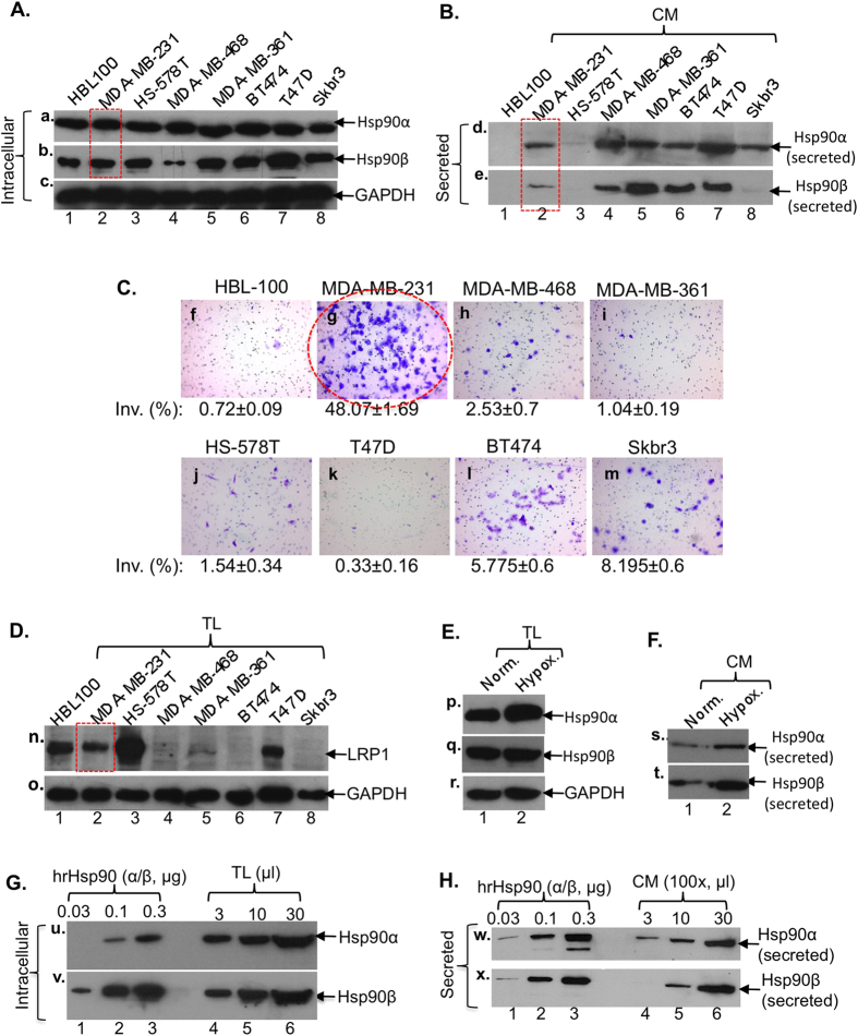
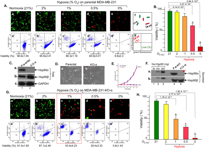
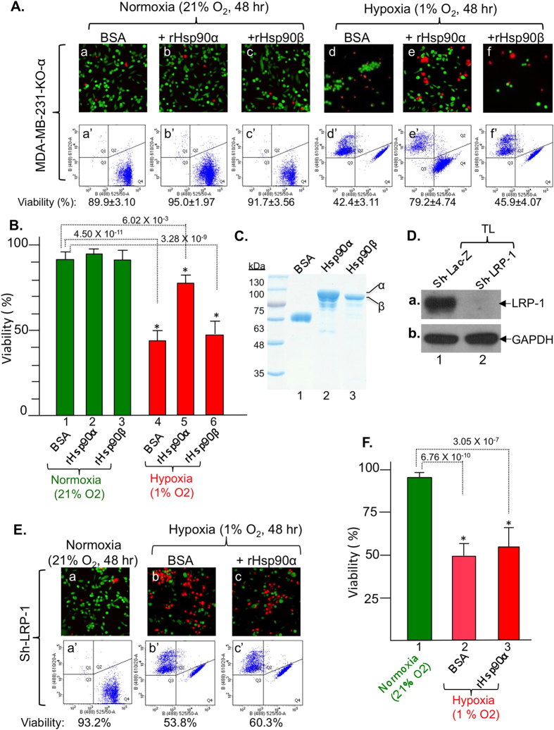
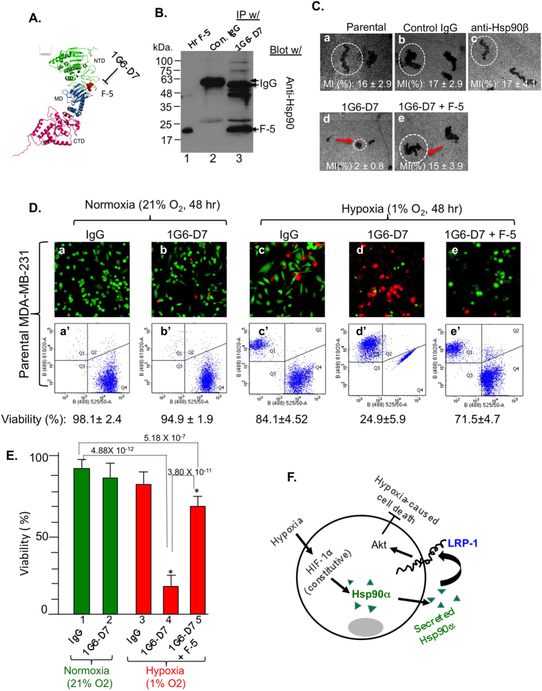
Rapidly growing tumours in vivo often outgrow their surrounding available blood supply, subjecting themselves to a severely hypoxic microenvironment. Understanding how tumour cells adapt themselves to survive hypoxia may help to develop new treatments of the tumours. Given the limited blood perfusion to the enlarging tumour, whatever factor(s) that allows the tumour cells to survive likely comes from the tumour cells themselves or its associated stromal cells. In this report, we show that HIF-1α-overexpressing breast cancer cells, MDA-MB-231, secrete heat shock protein-90alpha (Hsp90α) and use it to survive under hypoxia. Depletion of Hsp90α secretion from the tumour cells was permissive to cytotoxicity by hypoxia, whereas supplementation of Hsp90α-knockout tumour cells with recombinant Hsp90α, but not Hsp90β, protein prevented hypoxia-induced cell death via an autocrine mechanism through the LDL receptor-related protein-1 (LRP1) receptor. Finally, direct inhibition of the secreted Hsp90α with monoclonal antibody, 1G6-D7, enhanced tumour cell death under hypoxia. Therefore, secreted Hsp90α is a novel survival factor for certain tumours under hypoxia.


Figures

Figure 1
Figure 1. Selection of MDA-MB-231 breast cancer cell line as the model of study.
Western blots for Hsp90α and Hsp90β in total lysates (TL) (A) or conditioned media (100×) (CM) (B) and the invasiveness (C) of the indicated breast cancer and control cell lines. Western blots for LRP-1 receptor among cell lines (D) and the total (E) and the secreted (F) Hsp90α and Hsp90β under hypoxia in MDA-MB-231. Intracellular (G) and secreted (H) ratios between Hsp90α and Hsp90β in MDA-MB-231 cells.
Figure 2
Figure 2. CRISPR-cas9 knockout of Hsp90α sensitizes MDA-MB-231 cells to hypoxia-driven killing.
(A) Cell viability by fluorescence microscopy (panels a to e) and flow cytometry (panels a’ to e’). (B) Quantitation of viability data. (C) Evidence of CRISPR/Cas9 knockout of Hsp90α protein, (D) morphology and (E) proliferation profiles of parental and Hsp90α-knockout cells. (F) Depletion of Hsp90α (panel a), but not Hsp90β (panel b), secretion in Hsp90α-knockout cells. (G) Viability of Hsp90α-knockout cells under normoxia or various degrees of hypoxia (H) Quantitation of viability data. n = 3, *p < 0.05.
Figure 3
Figure 3. Rescue of Hsp90α-knockout cells from hypoxia-driven killing by extracellular Hsp90α, but not Hsp90β, protein via LRP-1 receptor signaling.
(A) Extracellular Hsp90α, not Hsp90β, rescues viability of Hsp90α-knockout cells. (B) Quantitation of viability data. (C) Evidence of purified recombinant proteins for rescues. (D) Down-regulation of LRP-1 shown by Western blot. (E) No rescue of LRP-1-downregulated cells from hypoxia by extracellular Hsp90α. (F) Quantitation. n = 3, *p < 0.05.
Figure 4
Figure 4. mAb 1G6-D7 neutralizes secreted Hsp90α and sensitizes MDA-MB-231 cells to hypoxia-driven killing.
(A) A schematic location for the F-5 in Hsp90α and 1G6-D7’s recognition. (B) mAb 1G6-D7 immunoprecipitates purified native F-5 protein. (C) mAb 1G6-D7 (10 μg/ml) blocked MDA-MB-231 cell migration (panel d) and F-5 (30 μg/ml) reversed the inhibition (panel e). (D) 1G6-D7 (panels d and d’) causes increased cell death under hypoxia and F-5 reversed 1G6-D7 effect. (E) Quantitation, n = 3, *p < 0.05. (F). A possible new mechanism for tumour cells’ survival under hypoxia: the HIF-1 > Hsp90α secretion > LRP1 receptor > Akt pathway.

References

    1. Bertout J. A., Patel S. A. & Simon M. C. The impact of O2 availability on human cancer. Nat Rev Cancer 8, 967–975 (2008). - PMC - PubMed
    1. Semenza G. L. Hypoxia-inducible factors in physiology and medicine. Cell 148, 399–408 (2012). - PMC - PubMed
    1. Majmundar A. J., Wong W. J. & Simon M. C. Hypoxia-inducible factors and the response to hypoxic stress. Mol Cell 40, 294–309 (2010). - PMC - PubMed
    1. Dales J. P. et al. Overexpression of hypoxia-inducible factor HIF-1alpha predicts early relapse in breast cancer: retrospective study in a series of 745 patients. Int. J. Cancer 116, 734–739 (2005). - PubMed
    1. Lundgren K., Holm C. & Landberg G. Hypoxia and breast cancer: prognostic and therapeutic implications. Cell Mol Life Sci. 64, 3233–3247 (2007). - PMC - PubMed

Publication types

Substances