. 2016 Apr;29(4):421-6.
doi: 10.1038/modpathol.2016.32. Epub 2016 Feb 5.

THSD7A staining of membranous glomerulopathy in clinical practice reveals cases with dual autoantibody positivity

Affiliations

THSD7A staining of membranous glomerulopathy in clinical practice reveals cases with dual autoantibody positivity

Christopher P Larsen et al. Mod Pathol. 2016 Apr.

Abstract

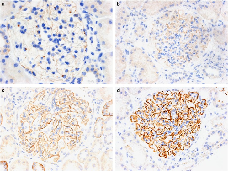
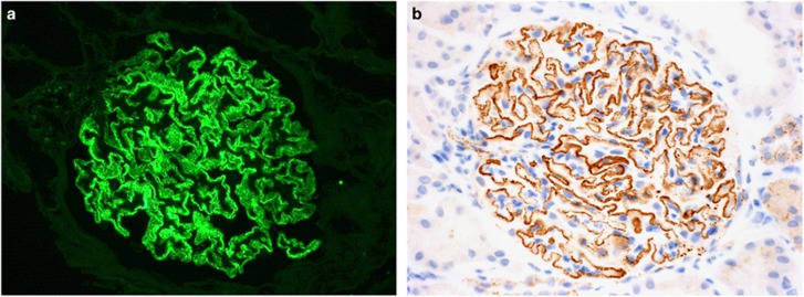
Thrombospondin type I domain-containing 7A (THSD7A) is a known antigenic target of autoantibodies leading to primary membranous glomerulopathy and was reported to account for ~10% of phospholipase A2 receptor (PLA2R)-negative membranous glomerulopathy. It has been proposed that PLA2R and THSD7A autoantibodies are mutually exclusive in membranous glomerulopathy. We validated an immunohistochemical assay to investigate for THSD7A-associated membranous glomerulopathy and utilized it in 258 consecutive native kidney biopsies, which showed membranous glomerulopathy in our laboratory, with the exception of membranous lupus nephritis. Membranous glomerulopathy stained positive for THSD7A-only in 7 (3%) cases, PLA2R-only in 141 (55%) cases, and showed dual positivity for THSD7A and PLA2R in 2 (1%) cases. Serologic testing for antibodies to PLA2R and THSD7A was performed in a subset of these patients. There was 100% correlation between positive THSD7A and/or PLA2R tissue staining and the presence of the corresponding autoantibodies in the serum including the two cases with dual positive THSD7A and PLA2R antibodies. We describe and provide a protocol for detection of THSD7A-associated membranous glomerulopathy in clinical practice. The cases with dual THSD7A and PLA2R positivity show that these autoantibodies are not mutually exclusive. They also emphasize the importance of using a panel-based approach when subtyping membranous glomerulopathy as a patient could conceptually be identified and treated based on anti-PLA2R titers, but still have anti-THSD7A antibodies driving persistent disease.


Conflict of interest statement

CPL and LNC declare no competing interests. LHB is a co-inventor on the US patent ‘Diagnostics for Membranous Nephropathy'.

Figures

Figure 1
Figure 1
Immunoperoxidase staining for THSD7A in membranous glomerulopathy. (a) THSD7A staining in a normal glomerulus shows weak segmental staining of the glomerular basement membranes. Original magnification × 400. (b and c): Glomeruli from two different cases of PLA2R-associated membranous glomerulopathy demonstrate the range of normal THSD7A staining that can be seen in cases of membranous glomerulopathy from (b) weak segmental staining along the glomerular basement membranes to (c) a global moderate pseudo-linear pattern. Original magnification × 400. (d) Strong global granular capillary loop staining is evident in this case of THASD7A-associated membranous glomerulopathy. Original magnification × 400.
Figure 2
Figure 2
Membranous glomerulopathy with dual positive staining. (a and b) This case of membranous glomerulopathy showed strong global granular capillary wall staining for both (a) PLA2R (indirect immunofluorescence; original magnification × 400) and (b) THSD7A (immunoperoxidase; original magnification × 400).
Figure 3
Figure 3
Western blotting with patient serum to detect reactivity against PLA2R and THSD7A. (a) Sera were initially screened by Western blotting against human glomerular extract (HGE) and cell-expressed recombinant PLA2R. Several sera (1 and 3) react with a band in HGE that did not appear to be PLA2R. (b) Representative sera are blotted against both recombinant PLA2R and THSD7A. For case 1 (and case 3, not shown) the reactivity corresponding to the band seen in HGE in panel A is confirmed to be against THSD7A. Immunoblotting against both antigens shows that case 4 has reactivity to both PLA2R and THSD7A. Repeated use of a very limited amount of the initial serum sample explains the weak PLA2R signal for case 4. A follow-up (4f/u) serum was obtained that clearly shows reactivity with both antigens. (c) To confirm reactivity with PLA2R, sera were additionally tested by Western blot against the immunodominant epitope, which resides in CysR, as well as a minor epitope in CTLD1. Although none of the cases had reactivity to CTLD1, the anti-PLA2R only cases (2 and 5) and the dual anti-PLA2R and anti-THSD7A case (4) exhibited reactivity with the CysR domain of PLA2R. P-T+, anti-PLA2R negative and anti-THSD7A positive; P+T-, anti-PLA2R positive and anti-THSD7A negative; P+T+ dual positive for anti-PLA2R and anti-THSD7A.

References

    1. Beck LH, Bonegio RG, Lambeau G et al. M-type phospholipase A2 receptor as target antigen in idiopathic membranous nephropathy. N Engl J Med 2009;361:11–21. - PMC - PubMed
    1. Larsen CP, Messias NC, Silva FG et al. Determination of primary versus secondary membranous glomerulopathy utilizing phospholipase A2 receptor (PLA2R1) staining in renal biopsies. Mod Pathol 2013;26:709–715. - PubMed
    1. Hoxha E, Harendza S, Zahner G et al. An immunofluorescence test for phospholipase-A2-receptor antibodies and its clinical usefulness in patients with membranous glomerulonephritis. Nephrol Dial Transplant 2011;26:2526–2532. - PubMed
    1. Ruggenenti P, Debiec H, Ruggiero B et al. Anti-phospholipase A2 receptor antibody titer predicts post-rituximab outcome of membranous nephropathy. J Am Soc Nephrol 2015;26:2545–2458. - PMC - PubMed
    1. Hoxha E, Thiele I, Zahner G et al. Phospholipase A2 receptor autoantibiodies and clinical outcome in patients with primary membranous nephropathy. J Am Soc Nephrol 2014;25:1357–1366. - PMC - PubMed

Publication types

LinkOut - more resources