. 2016 Feb 5:4:10.
doi: 10.1186/s40478-016-0284-9.

Emergence of two prion subtypes in ovine PrP transgenic mice infected with human MM2-cortical Creutzfeldt-Jakob disease prions

Affiliations

Emergence of two prion subtypes in ovine PrP transgenic mice infected with human MM2-cortical Creutzfeldt-Jakob disease prions

Jérôme Chapuis et al. Acta Neuropathol Commun. .

Abstract

Introduction: Mammalian prions are proteinaceous pathogens responsible for a broad range of fatal neurodegenerative diseases in humans and animals. These diseases can occur spontaneously, such as Creutzfeldt-Jakob disease (CJD) in humans, or be acquired or inherited. Prions are primarily formed of macromolecular assemblies of the disease-associated prion protein PrP(Sc), a misfolded isoform of the host-encoded prion protein PrP(C). Within defined host-species, prions can exist as conformational variants or strains. Based on both the M/V polymorphism at codon 129 of PrP and the electrophoretic signature of PrP(Sc) in the brain, sporadic CJD is classified in different subtypes, which may encode different strains. A transmission barrier, the mechanism of which remains unknown, limits prion cross-species propagation. To adapt to the new host, prions have the capacity to 'mutate' conformationally, leading to the emergence of a variant with new biological properties. Here, we transmitted experimentally one rare subtype of human CJD, designated cortical MM2 (129 MM with type 2 PrP(Sc)), to transgenic mice overexpressing either human or the VRQ allele of ovine PrP(C).

Results: In marked contrast with the reported absence of transmission to knock-in mice expressing physiological levels of human PrP, this subtype transmitted faithfully to mice overexpressing human PrP, and exhibited unique strain features. Onto the ovine PrP sequence, the cortical MM2 subtype abruptly evolved on second passage, thereby allowing emergence of a pair of strain variants with distinct PrP(Sc) biochemical characteristics and differing tropism for the central and lymphoid tissues. These two strain components exhibited remarkably distinct replicative properties in cell-free amplification assay, allowing the 'physical' cloning of the minor, lymphotropic component, and subsequent isolation in ovine PrP mice and RK13 cells.

Conclusions: Here, we provide in-depth assessment of the transmissibility and evolution of one rare subtype of sporadic CJD upon homologous and heterologous transmission. The notion that the environment or matrix where replication is occurring is key to the selection and preferential amplification of prion substrain components raises new questions on the determinants of prion replication within and between species. These data also further interrogate on the interplay between animal and human prions.


Figures
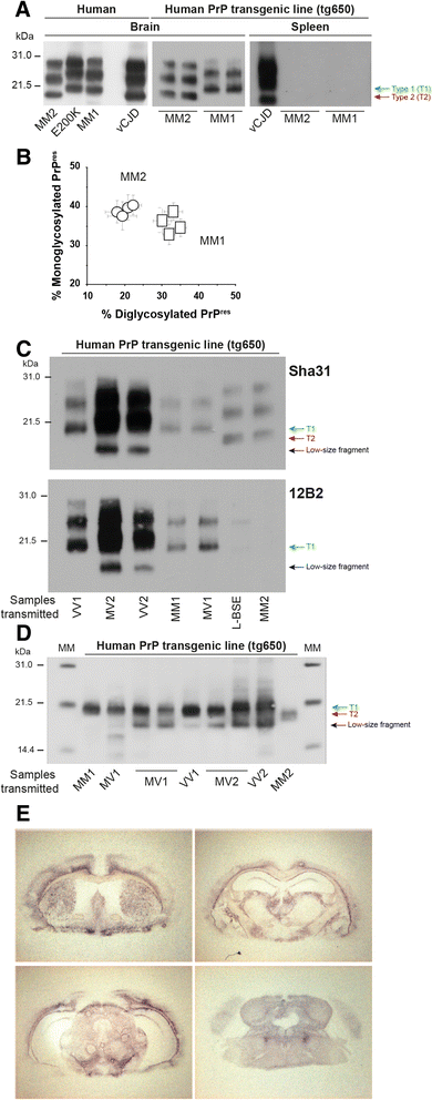
Fig. 1
Fig. 1
Biochemical and histopathological strain phenotype of MM2-sCJD prions in human PrP mice. (a) Electrophoretic pattern of cortical MM2-sCJD prions in human brain and in human PrP mouse (tg650) brains and spleens. Tissue homogenates were subjected to western blot analyses after limited proteinase K digestion. Blots were probed with Sha31 antibody. Other human prion sources (MM1-sCJD, E200K familial CJD and variant CJD) are shown as controls. The equivalent of 0.5 and 2 mg of brain and spleen tissue was loaded on the SDS-PAGE gel. Red and blue arrows denote the unglysosylated bands of PrPres migrating at 21 kDa (T1) and 19 kDa (T2), respectively. Molecular masses (MM) of protein standards are indicated in kilodaltons. (b) Ratio of diglycosylated and monoglycosylated PrPres species in the brains of tg650 mice following serial transmission (4 passages) of MM2-sCJD (circles) and MM1-sCJD (squares) prions (data plotted as means ± SEM, n = 6 mice analyzed at each passage). (c) Western blot analysis of PrPres in the brain of tg650 mice infected with MM2-sCJD prions, after blotting with Sha31 antibody (top) or 12B2 antibody specific for Type 1 PrPres (bottom). The banding patterns observed on transmission of other CJD subtypes and atypical L-BSE (which exhibits also a T2 signature, [4]) are shown for comparison. The equivalent of 1 mg (Sha31) and 7 mg (12B12) tissue were run on the SDS-PAGE gels for MM2-sCJD and L-BSE infected brains. The equivalent of 1 mg (Sha31, VV1, MM1, MV1; 12B2, MV2, VV2), 0.5 mg (Sha31 MV2, VV2), 2 mg (12B2, VV1, MM1, MV1) were loaded for the other samples. Note the presence of low-size PrPres fragments in the brain of tg650 mice infected with MV2 and VV2 sCJD sources (black arrow). (d) Western blot analysis of PrPres in the brain of tg650 mice infected with MM2-sCJD prions, after deglycosylation by PNGase F. Blots were probed with Sha31. Other CJD subtypes are shown for comparison. The equivalent of 0.2 mg of brain tissue (MM1, MM2), 0.1 mg (MV1, VV1) and 0.05 mg (MV2, VV2) were run on the SDS-PAGE gels. MM: molecular mass standards. (e) Regional distribution of PrPres in the brain of tg650 mice infected with MM2-sCJD prions, by representative histoblots in 4 different antero-posterior sections. See [7] for comparison with transmission of MM1-sCJD prions. Histoblots were probed with 12F10 anti-PrP antibody
Fig. 2
Fig. 2
Banding patterns of PrPres in the brain and spleen tissue of ovine PrP mice infected with MM2-sCJD prions. (a) PrPres banding patterns in the brain and spleen tissue of ovine PrP mice (tg338 line) over two serial passages of MM2-sCJD (MM2). Note the different patterns in brain and spleen, which were designated T2Ov (red arrow) and T1Ov (blue arrow), respectively. These patterns can be compared with those observed after inoculation of 127S scrapie prions, a strain with a 21 kDa signature in brain and spleen [34]. (b) Immunoblots showing PrPres pattern in the brain and spleen of tg338 mice over serial passage of uncloned MM2-sCJD (MM2) and cloned (Cl.) MM2-sCJD prions. Note the low levels of T2Ov PrPres in the spleens of tg338 mice inoculated with cloned MM2-sCJD prions, which are only visible on overexposed gels. The equivalent of 1 and 2 mg of tg338 brain and spleen tissue was loaded on the SDS-PAGE gels. The blots were probed with Sha31 antibody
Fig. 3
Fig. 3
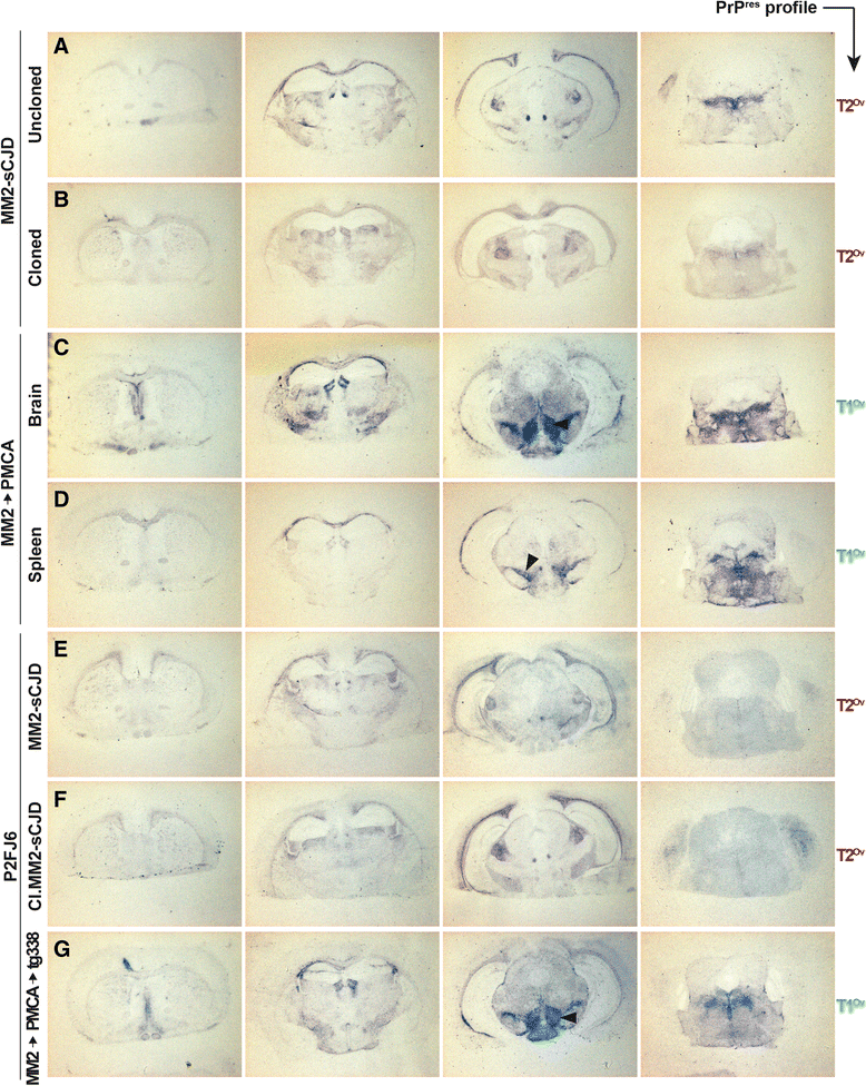
PrPres deposition pattern in the brain of ovine PrP mice challenged with MM2-sCJD prions. Representative histoblots in 4 different antero-posterior sections of tg338 mouse brain after inoculation with tg338-passaged MM2-sCJD prions (a, 4th passage), tg338-cloned MM2-sCJD prions (b), brain (c) and spleen (d) of tg338 mice inoculated with PMCA-amplified MM2-sCJD prions, P2FJ6 cells challenged with uncloned (e), cloned (f) and PMCA-derived (g) MM2-sCJD prions (MM2 → PMCA → tg338). Note the marked deposition of PrPres in the lateral hypothalamic areas of mice inoculated with T1Ov prions (black arrowhead). Blots were probed with 12F10
Fig. 4
Fig. 4
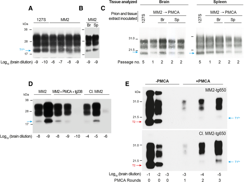
Amplification of MM2-sCJD prions by PMCA, using ovine PrPC as substrate. (a, b) Endpoint titration of tg338-passaged MM2-sCJD prions by a single round of PMCA. Brain (a) or spleen (b) homogenates from tg338 mice infected with MM2-sCJD prions were serially diluted in tg338 healthy brain lysate, as indicated, and submitted to one round of PMCA. Each amplified dilution was analyzed by immunoblotting (Sha31 antibody) for the presence of PrPres and characterization of electrophoretic pattern. Amplification of 127S prion seeds (21 kDa-PrPres) has been done in parallel for comparison [41]. (c) PrPres banding pattern in the brain and spleen tissue of tg338 mice inoculated with PMCA amplified products from tg338-passaged MM2-sCJD prions (10−8 dilution seed) and on subpassage (MM2 → PMCA). On 2nd passage, mice were challenged with either brain (Br) or spleen (Sp) extracts. The equivalent of 1 and 2 mg of brain and spleen tissue was loaded on the SDS-PAGE gels (Sha31 antibody). 127S PrPres is shown as control. (d) Endpoint titration by PMCA of uncloned (MM2) and cloned (Cl.MM2) tg338-passaged MM2-sCJD prions. The limiting dilution achieved was compared to that observed after PMCA reactions seeded with brain homogenates from tg338 mice inoculated with PMCA-amplified MM2-sCJD prions (2nd passage; MM2 → PMCA → tg338). (e) Amplification of human MM2-sCJD prions by PMCA, using tg338 healthy brain lysate as substrate. PMCA reactions were seeded with 103-diluted brain material from human PrP tg650 mice infected with uncloned (3rd passage; MM2-tg650) or cloned (4th passage; Cl.MM2-tg650) Three amplification rounds were performed. After each round, the amplified products were analyzed for PrPres content and electrophoretic pattern by immunoblot (Sha31 antibody) or diluted 1:10 in fresh substrate to seed the following round
Fig. 5
Fig. 5
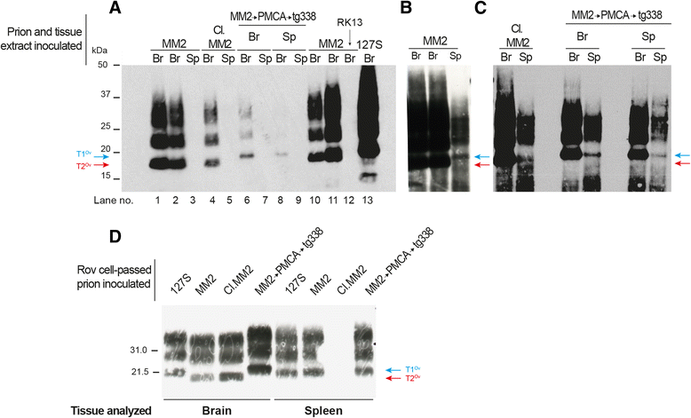
Infection of P2FJ6 cells with ovine MM2-SCJD prions, and PrPres glycopattern of the cell-passaged prions in tg338 mice. (a-c) Western blot analysis of PrPres in P2FJ6 cell lysates infected with brain (Br) and spleen (Sp) extracts of tg338 mice infected with MM2-sCJD prions, either uncloned (MM2) or cloned (Cl.MM2), and mice challenged with either brain or spleen extracts of PMCA MM2-sCJD prions (MM2 → PMCA → tg338). The results are shown over 3 (a) or 4 passages (b, c) post-cell exposure, except in lanes 11-12 in (A), where the analysis was made at 14 passages. The gels in b and c are overexposed purposely to reveal accumulation of T1Ov PrPres after infection with spleen material. Untransfected RK13 cells (RK13, arrow in a) did not accumulate detectable levels of PrPres. Infection with 127S prions was performed in parallel to estimate the efficacy of infection. The gels were loaded with 250 μg of PK-digested protein or 80 μg for 127S infection. The blots were probed with Sha31 antibody. (d) Western blot analysis of PrPres in the brain and spleen tissue of tg338 mice challenged with P2FJ6 cell lysates infected with uncloned (MM2), cloned (Cl.MM2) MM2-sCJD prions, PMCA MM2-sCJD prions (MM2 → PMCA → tg338), and 127S as control. The equivalent of 1 and 2 mg of brain and spleen tissue was loaded on the SDS-PAGE gels. The blots were probed with Sha31 antibody

Similar articles

Cited by

References

    1. Andreoletti O, Orge L, Benestad SL, Beringue V, Litaise C, Simon S, et al. Atypical/Nor98 scrapie infectivity in sheep peripheral tissues. PLoS pathogens. 2011;7 doi: 10.1371/journal.ppat.1001285. - DOI - PMC - PubMed
    1. Angers RC, Kang HE, Napier D, Browning S, Seward T, Mathiason C, et al. Prion strain mutation determined by prion protein conformational compatibility and primary structure. Science. 2010;328:1154–1158. doi: 10.1126/science.1187107. - DOI - PMC - PubMed
    1. Barria MA, Telling GC, Gambetti P, Mastrianni JA, Soto C. Generation of a new form of human PrP(Sc) in vitro by interspecies transmission from cervid prions. The Journal of biological chemistry. 2011;286:7490–7495. doi: 10.1074/jbc.M110.198465. - DOI - PMC - PubMed
    1. Beringue V, Andreoletti O, Le Dur A, Essalmani R, Vilotte JL, Lacroux C, et al. A bovine prion acquires an epidemic bovine spongiform encephalopathy strain-like phenotype on interspecies transmission. J Neurosci. 2007;27:6965–6971. doi: 10.1523/JNEUROSCI.0693-07.2007. - DOI - PMC - PubMed
    1. Beringue V, Herzog L, Jaumain E, Reine F, Sibille P, Le Dur A, et al. Facilitated cross-species transmission of prions in extraneural tissue. Science. 2012;335:472–475. doi: 10.1126/science.1215659. - DOI - PubMed

Publication types

MeSH terms